How to draw a heart organ. Images and anatomical links
The heart means a lot to a person. A real heart is the basis of our body, and valentines or simple drawn hearts help us express our feelings. This is a manifestation of warmth, love and tender feelings to a person. Below we will give a few simple tips how to draw a heart. There are several drawing options, you can use them or come up with your own.
Simplified version
Before you draw a heart with a pencil (or rather, start), prepare all the tools (paper, eraser, pencils). Place a piece of paper in front of you. First you need to think through the details if you want to add something to the heart. Make sure that all parts of the drawing fit on the sheet. It is better to draw all the main elements schematically (in squares, circles). Now take a pencil and get started. There are three options for how to draw a Valentine heart

First way
Place a dot in the center of the sheet, it will be the base of the heart. Draw a semicircular line, directing it first up to the right and then down. The end point of the arc should be under the base point. You should end up with something that looks like a question mark. Repeat the steps on the left half of the sheet. The lines should converge at one point.
Second way
Draw upside down isosceles triangle(the base should be at the top). Draw a bisector from the bottom vertex. Then “write” half a heart into each of the resulting triangles. Use an eraser to remove unnecessary lines.
Third way
Draw two intersecting circles (you can use stencils) and draw a heart based on them. If it turns out to be asymmetrical, then fold a sheet of paper in half and draw one half at the fold line, then cut it out. Now you know how to draw beautiful heart in its simplified version. When you have the base ready, you can use your imagination: pierce the heart with arrows, thorns, draw roses or wings around it. You can color it or outline it with a marker, leaving it in black and white. Do not overload the drawing with many unnecessary details.
How to draw a human heart
You will also need tools, prepare yourself a space. It is better to use a vertically oriented sheet. In this matter, you need to thoroughly study the anatomy of the human heart. You can copy it from a textbook or medical reference book.

Brief description of the process:
You need to draw an oval that tapers downwards. It should be slightly tilted. Then draw the right atrium. An important part of the heart is the aorta, don’t forget about it. This is a large “tube” that will be located at the top of the picture, with three more vessels coming out of it. Add the veins, don't forget the left atrium. Also trace the drawing and color it if desired. Don't forget to erase extra lines.
Conclusion
Now you know several ways to draw a heart. If you don't get the drawing, don't give up. When everything starts to work out for you, you can please your loved one with a beautiful handmade valentine.
Anatomy and physiology of the heart: structure, functions, hemodynamics, cardiac cycle, morphology
The structure of the heart of any organism has many characteristic nuances. In the process of phylogenesis, that is, the evolution of living organisms to more complex ones, the heart of birds, animals and humans acquires four chambers instead of two chambers in fish and three chambers in amphibians. Such a complex structure the best way adapted to separate arterial and venous blood flows. In addition, the anatomy of the human heart involves many the smallest details, each of which performs its strictly defined functions.
Heart as an organ
So, the heart is nothing more than a hollow organ consisting of specific muscle tissue, which carries out the motor function. The heart is located in the chest behind the sternum, more to the left, and its longitudinal axis is directed anteriorly, to the left and down. In front, the heart borders on the lungs, almost completely covering them, leaving only a small part directly adjacent to the chest from the inside. The boundaries of this part are otherwise called absolute cardiac dullness, and they can be determined by tapping the chest wall ().
In people with a normal constitution, the heart has a semi-horizontal position in the chest cavity, in people with an asthenic constitution (thin and tall) it is almost vertical, and in hypersthenics (dense, stocky, with a large muscle mass) – almost horizontal.

heart position
The posterior wall of the heart is adjacent to the esophagus and to the large main vessels (thoracic aorta, inferior vena cava). The lower part of the heart is located on the diaphragm.

external structure of the heart
Age characteristics
The human heart begins to form in the third week of the intrauterine period and continues throughout the entire period of gestation, passing through stages from a single-chamber cavity to a four-chamber heart.

development of the heart in utero
The formation of four chambers (two atria and two ventricles) occurs already in the first two months of pregnancy. The smallest structures are fully formed by birth. It is in the first two months that the embryonic heart is most vulnerable to negative influence some factors on the expectant mother.
The fetal heart participates in the blood flow throughout its body, but differs in the circles of blood circulation - the fetus does not yet have its own breathing with lungs, and it “breathes” through placental blood. There are some openings in the fetal heart that allow pulmonary blood flow to be “switched off” from the circulation before birth. During childbirth, accompanied by the first cry of the newborn, and, consequently, at the moment of increased intrathoracic pressure and pressure in the baby's heart, these openings close. But this does not always happen, and the child may still have them, for example (not to be confused with a defect such as atrial septal defect). Open window is not a heart defect, and subsequently, as the child grows, it heals.

hemodynamics in the heart before and after birth
The heart of a newborn baby has a round shape, and its dimensions are 3-4 cm in length and 3-3.5 cm in width. In the first year of a child's life, the heart increases significantly in size, more in length than in width. The weight of a newborn baby's heart is about 25-30 grams.
As the baby grows and develops, the heart also grows, sometimes significantly ahead of the development of the body itself according to age. By the age of 15, the mass of the heart increases almost tenfold, and its volume increases more than fivefold. The heart grows most rapidly until the age of five, and then during puberty.
In an adult, the size of the heart is about 11-14 cm in length and 8-10 cm in width. Many people rightly believe that the size of each person’s heart corresponds to the size of his clenched fist. The weight of the heart in women is about 200 grams, and in men it is about 300-350 grams.
After age 25, changes begin in the connective tissue of the heart, which forms the heart valves. Their elasticity is no longer the same as in childhood and adolescence, and the edges may become uneven. As a person grows and then ages, changes occur in all structures of the heart, as well as in the vessels that feed it (the coronary arteries). These changes can lead to the development of numerous cardiac diseases.
Anatomical and functional features of the heart
Anatomically, the heart is an organ divided into four chambers by septa and valves. The “upper” two are called atria (atrium), and the “lower” two are called ventricles (ventriculum). Between the right and left atria is the interatrial septum, and between the ventricles is the interventricular septum. Normally, these septa do not have holes in them. If there are holes, this leads to mixing of arterial and venous blood, and, accordingly, to hypoxia of many organs and tissues. Such holes are called septal defects and are classified as.

basic structure of the chambers of the heart
The boundaries between the upper and lower chambers are the atrioventricular openings - the left one, covered by the mitral valve leaflets, and the right one, covered by the tricuspid valve leaflets. The integrity of the septa and the proper operation of the valve leaflets prevent the mixing of blood flows in the heart and promote clear unidirectional blood flow.
The atria and ventricles are different - the atria are smaller than the ventricles and have thinner walls. Thus, the wall of the atria is about only three millimeters, the wall of the right ventricle is about 0.5 cm, and the wall of the left is about 1.5 cm.
The atria have small projections called ears. They have a slight suction function for better pumping of blood into the atrium cavity. The mouth of the vena cava flows into the right atrium near its appendage, and four (less often five) pulmonary veins flow into the left atrium. The pulmonary artery (more often called the pulmonary trunk) on the right and the aortic bulb on the left depart from the ventricles.

structure of the heart and its vessels
From the inside, the upper and lower chambers of the heart are also different and have their own characteristics. The surface of the atria is smoother than the ventricles. Thin connective tissue valves originate from the valve ring between the atrium and the ventricle - bicuspid (mitral) on the left and tricuspid (tricuspid) on the right. The other edge of the valves faces the inside of the ventricles. But so that they do not hang freely, they are supported, as it were, by thin tendon threads called chords. They are like springs, stretch when the valve flaps close and compress when the valve flaps open. The chordae originate from the papillary muscles from the wall of the ventricles - three in the right and two in the left ventricle. That is why the ventricular cavity has an uneven and lumpy inner surface.
The functions of the atria and ventricles also differ. Due to the fact that the atria need to push blood into the ventricles, and not into larger and longer vessels, they have to overcome less resistance from muscle tissue, therefore the atria are smaller in size and their walls are thinner than those of the ventricles. The ventricles push blood into the aorta (left) and the pulmonary artery (right). Conventionally, the heart is divided into right and left halves. The right half serves for the flow of exclusively venous blood, and the left half for arterial blood. Schematically, the “right heart” is indicated in blue, and the “left heart” is indicated in red. Normally, these flows never mix.

hemodynamics in the heart
One cardiac cycle lasts about 1 second and is carried out as follows. At the moment the atria are filled with blood, their walls relax - atrial diastole occurs. The valves of the vena cava and pulmonary veins are open. The tricuspid and mitral valves are closed. Then the atrial walls tense and push blood into the ventricles, the tricuspid and mitral valves are open. At this moment, systole (contraction) of the atria and diastole (relaxation) of the ventricles occur. After the ventricles receive blood, the tricuspid and mitral valves close, and the aortic and pulmonary valves open. Next, the ventricles contract (ventricular systole), and the atria fill with blood again. The general diastole of the heart begins.

cardiac cycle
The main function of the heart is reduced to pumping, that is, to pushing a certain blood volume into the aorta with such pressure and speed that the blood is delivered to the most distant organs and to the smallest cells of the body. Moreover, arterial blood with a high content of oxygen and nutrients is pushed into the aorta, entering the left half of the heart from the vessels of the lungs (flows to the heart through the pulmonary veins).
Venous blood, low in oxygen and other substances, is collected from all cells and organs from the venous cava system, and flows into the right half of the heart from the superior and inferior vena cava. Next, venous blood is pushed from the right ventricle into the pulmonary artery, and then into the pulmonary vessels in order to carry out gas exchange in the alveoli of the lungs and to enrich it with oxygen. In the lungs, arterial blood collects in the pulmonary venules and veins, and again flows into the left side of the heart (the left atrium). And so the heart regularly pumps blood throughout the body at a frequency of 60-80 beats per minute. These processes are designated by the concept "Circles of Blood Circulation". There are two of them - small and large:
- Small circle includes the flow of venous blood from the right atrium through the tricuspid valve into the right ventricle - then into the pulmonary artery - then into the arteries of the lungs - oxygenation of blood in the pulmonary alveoli - flow of arterial blood into the smallest veins of the lungs - into the pulmonary veins - into the left atrium.
- Big circle includes the flow of arterial blood from the left atrium through the mitral valve into the left ventricle - through the aorta into the arterial bed of all organs - after gas exchange in tissues and organs, the blood becomes venous (with a high content of carbon dioxide instead of oxygen) - then into the venous bed of organs - into the hollow system veins - into the right atrium.

circulation circles
Video: cardiac anatomy and cardiac cycle briefly
Morphological features of the heart
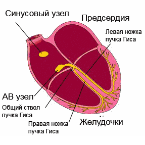
If you examine sections of the heart under a microscope, you can see a special type of muscle that is not found in any other organ. This is a type of striated muscle, but has significant histological differences from ordinary skeletal muscles and from the muscles lining internal organs. The main function of the heart muscle, or myocardium, is to provide most important ability the heart, which forms the basis of the vital activity of the entire organism as a whole. This is the ability to contract, or contractility.In order for the heart muscle fibers to contract synchronously, electrical signals must be supplied to them, which excite the fibers. This is another ability of the heart – .
Conduction and contractility are possible due to the fact that the heart autonomously generates electricity. Function data (automatism and excitability) are provided by special fibers that are integral part conducting system. The latter is represented by electrically active cells of the sinus node, atrioventricular node, the bundle of His (with two legs - right and left), as well as Purkinje fibers. In the case when a patient’s myocardial damage affects these fibers, they develop, otherwise called.
cardiac cycle
Normally, the electrical impulse originates in the cells of the sinus node, which is located in the area of the right atrium appendage. In a short period of time (about half a millisecond), the impulse spreads throughout the atrial myocardium and then enters the cells of the atrioventricular junction. Typically, signals are transmitted to the AV node through three main tracts - the Wenkenbach, Thorel and Bachmann bundles. In the cells of the AV node, the impulse transmission time is extended to 20-80 milliseconds, and then the impulses travel through the right and left branches (as well as the anterior and posterior branches of the left branch) of the His bundle to the Purkinje fibers, and ultimately to the working myocardium. The frequency of impulse transmission along all pathways is equal to the heart rate and is 55-80 impulses per minute.
So, the myocardium, or cardiac muscle, is the middle layer in the wall of the heart. The inner and outer membranes are connective tissue and are called endocardium and epicardium. The last layer is part of the pericardial sac, or cardiac “shirt”. Between the inner layer of the pericardium and the epicardium, a cavity is formed, filled with a very small amount of fluid, to ensure better sliding of the pericardial layers during heart contractions. Normally, the fluid volume is up to 50 ml; exceeding this volume may indicate pericarditis.

structure of the heart wall and membrane
Blood supply and innervation of the heart
Despite the fact that the heart is a pump to supply the entire body with oxygen and nutrients, it itself also needs arterial blood. In this regard, the entire wall of the heart has a well-developed arterial network, which is represented by the branching of the coronary (coronary) arteries. The orifices of the right and left coronary arteries depart from the root of the aorta and are divided into branches that penetrate the thickness of the heart wall. If these important arteries become clogged with blood clots and atherosclerotic plaques, the patient will develop and the organ will no longer be able to perform its functions fully.

location of the coronary arteries supplying blood to the heart muscle (myocardium)
The frequency and force with which the heart beats is influenced by nerve fibers extending from the most important nerve conductors - the vagus nerve and the sympathetic trunk. The first fibers have the ability to slow down the rhythm frequency, the latter - to increase the frequency and strength of the heartbeat, that is, they act like adrenaline.

innervation of the heart
In conclusion, it should be noted that the anatomy of the heart may have any deviations in individual patients, therefore, only a doctor can determine the norm or pathology in a person after conducting an examination that can most informatively visualize the cardiovascular system.
Video: lecture on cardiac anatomy
The heart is a muscular organ in humans and animals that pumps blood through blood vessels.
Functions of the heart - why do we need a heart?
Our blood provides the entire body with oxygen and nutrients. In addition, it also has a cleansing function, helping in the removal of metabolic waste.
The function of the heart is to pump blood through blood vessels.
How much blood does the human heart pump?
The human heart pumps from 7,000 to 10,000 liters of blood in one day. This amounts to approximately 3 million liters per year. That works out to 200 million liters over a lifetime!
The amount of blood pumped within a minute depends on the current physical and emotional load - the greater the load, the more blood the body requires. So the heart can conduct from 5 to 30 liters through itself in one minute.
The circulatory system consists of about 65 thousand vessels, their total length is about 100 thousand kilometers! Yes, we didn't make a mistake.
Circulatory system
The human cardiovascular system is formed by two circles of blood circulation. With each heartbeat, blood moves in both circles at once.
Pulmonary circulation
- Deoxygenated blood from the superior and inferior vena cava enters the right atrium and then into the right ventricle.
- From the right ventricle, blood is pushed into the pulmonary trunk. The pulmonary arteries carry blood directly to the lungs (to the pulmonary capillaries), where it receives oxygen and releases carbon dioxide.
- Having received enough oxygen, the blood returns to the left atrium of the heart through the pulmonary veins.
Systemic circulation
- From the left atrium, blood moves into the left ventricle, from where it is subsequently pumped through the aorta to big circle blood circulation
- After going through a difficult path, the blood again arrives through the vena cava to the right atrium of the heart.
Normally, the amount of blood pushed out of the ventricles of the heart is the same with each contraction. Thus, an equal volume of blood simultaneously enters the greater and lesser circulation.
What is the difference between veins and arteries?
- Veins are designed to transport blood to the heart, and the job of arteries is to supply blood in the opposite direction.
- In veins, blood pressure is lower than in arteries. Accordingly, the walls of arteries are more elastic and dense.
- Arteries saturate “fresh” tissue, and veins take away “waste” blood.
- In the case of vascular damage, arterial or venous bleeding can be distinguished by its intensity and the color of the blood. Arterial - strong, pulsating, beating like a “fountain”, the color of the blood is bright. Venous - bleeding of constant intensity (continuous flow), the color of the blood is dark.
The weight of a human heart is only about 300 grams (on average 250 grams for women and 330 grams for men). Despite the relatively low weight, this is undoubtedly main muscle in the human body and the basis of its life activity. The size of the heart is indeed approximately equal to a human fist. Athletes' hearts can be one and a half times larger than those of the average person.
Anatomical structure
The heart is located in the middle of the chest at the level of 5-8 vertebrae.
Normally, the lower part of the heart is located for the most part in the left half of the chest. There is a variant of congenital pathology in which all organs are mirrored. It's called transposition internal organs. The lung, next to which the heart is located (normally the left one), is smaller in size relative to the other half.
The back surface of the heart is located near the spinal column, and the front surface is reliably protected by the sternum and ribs.
The human heart consists of four independent cavities (chambers) divided by partitions:
- two upper ones - the left and right atria;
- and two lower ones - the left and right ventricles.
The right side of the heart includes the right atrium and ventricle. The left half of the heart is represented, respectively, by the left ventricle and atrium.
The inferior and superior vena cava enter the right atrium, and the pulmonary veins enter the left atrium. From right ventricle the pulmonary arteries (also called the pulmonary trunk) emerge. From left ventricle the ascending aorta rises.
The heart has protection from overstretching and other organs, which is called the pericardium or pericardial sac (a kind of membrane in which the organ is enclosed). It has two layers: an outer dense, durable connective tissue called fibrous membrane of the pericardium and internal ( serous pericardium).
Thus, the heart itself consists of three layers: epicardium, myocardium, endocardium. It is the contraction of the myocardium that pumps blood through the vessels of the body.
The walls of the left ventricle are approximately three times larger than the walls of the right! Explained this fact in that the function of the left ventricle is to push blood into the systemic circulation, where the resistance and pressure are much higher than in the pulmonary circulation.
The device of heart valves
Special heart valves allow you to constantly maintain blood flow in the correct (unidirectional) direction. The valves alternately open and close, either letting blood through or blocking its path. Interestingly, all four valves are located along the same plane.
Between the right atrium and the right ventricle is located tricuspid (tricuspid) valve. It contains three special leaflet plates that, during contraction of the right ventricle, can provide protection from the reverse flow (regurgitation) of blood into the atrium.
Works in a similar way mitral valve, only it is located on the left side of the heart and is bicuspid in its structure.
Aortic valve prevents the reverse flow of blood from the aorta into the left ventricle. Interestingly, when the left ventricle contracts, the aortic valve opens as a result of blood pressure on it, so it moves into the aorta. After which, during diastole (the period of relaxation of the heart), the reverse flow of blood from the artery promotes the closure of the valves.
Normally, the aortic valve has three leaflets. The most common congenital heart abnormality is bicuspid aortic valve. This pathology occurs in 2% of the human population.
Pulmonary valve at the moment of contraction of the right ventricle, it allows blood to flow into the pulmonary trunk, and during diastole it does not allow it to flow in the opposite direction. It also consists of three doors.
Cardiac vessels and coronary circulation
 The human heart requires nutrition and oxygen, just like any other organ. The vessels that supply (nourish) the heart with blood are called coronary or coronary. These vessels branch from the base of the aorta.
The coronary arteries supply the heart with blood, and the coronary veins remove deoxygenated blood. Those arteries that are located on the surface of the heart are called epicardial. Subendocardial arteries are called coronary arteries hidden deep in the myocardium.
Most of the blood outflow from the myocardium occurs through three cardiac veins: large, middle and small. Forming the coronary sinus, they flow into the right atrium. The anterior and small veins of the heart deliver blood directly to the right atrium.
Coronary arteries are divided into two types - right and left. The latter consists of the anterior interventricular and circumflex arteries. The great cardiac vein branches into the posterior, middle and small veins of the heart.
Even absolutely healthy people have their own unique features coronary circulation. In reality, the vessels may look and be located differently than shown in the picture.
How does the heart develop (form)?
Pulse path
This system ensures automatism of the heart - excitation of impulses generated in cardiomyocytes without an external stimulus. In a healthy heart main source impulses - sinoatrial (sinus) node. He is the leader and blocks the impulses from all other pacemakers. But if any disease occurs that leads to sick sinus syndrome, then other parts of the heart take over its function. Thus, the atrioventricular node (automatic center of the second order) and the His bundle (AC of the third order) are able to activate when the sinus node is weak. There are cases when secondary nodes enhance their own automaticity even during normal operation of the sinus node.
Sinus node located in the superior posterior wall of the right atrium in close proximity from the mouth of the superior vena cava. This node initiates pulses with a frequency of approximately 80-100 times per minute.
Atrioventricular node (AV) located in the lower part of the right atrium in the atrioventricular septum. This septum prevents the impulse from propagating directly into the ventricles, bypassing the AV node. If the sinus node is weakened, then the atrioventricular node will take over its function and begin to transmit impulses to the heart muscle at a frequency of 40-60 contractions per minute.
Next, the atrioventricular node passes into His bundle(atrioventricular bundle divided into two legs). The right leg rushes towards the right ventricle. The left leg is divided into two more halves.
The situation with the left bundle branch has not been fully studied. It is believed that the left leg with fibers from the anterior branch rushes to the anterior and lateral wall of the left ventricle, and the posterior branch supplies fibers to the posterior wall of the left ventricle and the lower parts of the lateral wall.
In case of weakness of the sinus node and atrioventricular block, the His bundle is capable of creating impulses at a speed of 30-40 per minute.
The conducting system deepens and further branches into smaller branches, eventually moving into Purkinje fibers, which penetrate the entire myocardium and serve as a transmission mechanism for contraction of the ventricular muscles. Purkinje fibers are capable of initiating impulses at a frequency of 15-20 per minute.
Exceptionally trained athletes can have normal resting heart rates down to the lowest recorded figure of just 28 beats per minute! However, for the average person, even one leading a very active lifestyle, a heart rate below 50 beats per minute may be a sign of bradycardia. If your heart rate is this low, you should be examined by a cardiologist.
Heartbeat
A newborn's heart rate may be around 120 beats per minute. With growing up the pulse ordinary person stabilizes between 60 and 100 beats per minute. Well trained athletes we're talking about about people with well-trained cardiovascular and respiratory systems) have a pulse of 40 to 100 beats per minute.
Controls heart rhythm nervous system- the sympathetic strengthens contractions, and the parasympathetic weakens.
Cardiac activity, to a certain extent, depends on the content of calcium and potassium ions in the blood. Other biologically active substances also contribute to the regulation of heart rhythm. Our heart may begin to beat faster under the influence of endorphins and hormones released when listening to our favorite music or kissing.
In addition, the endocrine system can have a significant impact on the heart rhythm - both the frequency of contractions and their strength. For example, the release of the well-known adrenaline by the adrenal glands causes an increase in heart rate. The hormone with the opposite effect is acetylcholine.
Heart sounds
One of the most simple methods diagnosing heart disease is by listening to the chest using a stethoscope (auscultation).
In a healthy heart, during standard auscultation, only two heart sounds are heard - they are called S1 and S2:
- S1 is the sound heard when the atrioventricular (mitral and tricuspid) valves close during ventricular systole (contraction).
- S2 - the sound heard when the semilunar (aortic and pulmonary) valves close during diastole (relaxation) of the ventricles.
Each sound consists of two components, but to the human ear they merge into one due to the very short period of time between them. If, under normal conditions of auscultation, additional tones become audible, this may indicate some kind of disease of the cardiovascular system.
Sometimes additional abnormal sounds may be heard in the heart, called a heart murmur. As a rule, the presence of murmurs indicates some kind of heart pathology. For example, noise can cause blood to flow back in the opposite direction (regurgitation) due to malfunction or damage to a valve. However, noise is not always a symptom of a disease. To clarify the reasons for the appearance of additional sounds in the heart, it is worth doing echocardiography (ultrasound of the heart).
Heart diseases
It is not surprising that the number of cardiovascular diseases. The heart is a complex organ that actually rests (if it can be called rest) only in the intervals between heartbeats. Any complex and constantly working mechanism itself requires as much as possible careful attitude and ongoing prevention.
Just imagine what a monstrous burden falls on the heart given our lifestyle and low-quality, abundant nutrition. Interestingly, mortality from cardiovascular diseases is quite high in countries with high level income.
The huge amounts of food consumed by the population of wealthy countries and the endless pursuit of money, as well as the associated stress, destroy our hearts. Another reason for the spread of cardiovascular diseases is physical inactivity - catastrophically low physical activity, destroying the entire body. Or, on the contrary, an illiterate passion for heavy physical exercise, often occurring in the background, the presence of which people do not even suspect and manage to die right during “health” activities.
Lifestyle and heart health
The main factors that increase the risk of developing cardiovascular diseases are:
- Obesity.
- High blood pressure.
- Increased blood cholesterol levels.
- Physical inactivity or excessive physical activity.
- Abundant, low-quality food.
- Depressed emotional condition and stress.
Do read this great article turning point in your life - give up bad habits and change your lifestyle.
Do you want to know how to draw human heart pencil step by step, take a few simple steps.
Step 1. Okay let's start this lesson on the human heart shall we? First draw out some guidelines and shapes so we have a nice workable wireframe to use. Start with a circle for the heart and then draw the bottom of the heart which contains the heart muscle. Three horizontal lines you see drawn there will be plugs on the pulmonary artery, pulmonary veins and left atrium. The lump you see drawn will be for the aorta.

Step 2. Okay, let's draw the actual shape of the aorta, as well as the pipes that come from this part of the heart. You will then draw out the shape for the pulmonary artery like you see here.

Step 3. Now, sketch out the shape of the vena cava that is in the vase while looking at the tube, which is all self-explanatory. The next draw is four pipes. The top tube enters the pulmonary artery, and the last three are the pulmonary veins, which are on the left side. Next sketch out the outer shape of the heart on the left side, which is also part of the heart muscle, and then draw in the veins that lie on the surface of the heart. Finally you will draw the tube for the inferior vena cava, which is located just below the lower left side of the heart.

Step 4. This is your last drawing step and all you need to do is draw out the remaining actual shape of the human heart and then draw in the superficial veins. Lastly, draw the tubes for the pulmonary vein and left atrium. Erase all the guidelines that are visible to clear your drawing of the human heart.