Description of bacteriophages. Bacteriophages
Bacteriophages These are viruses that selectively infect bacterial cells. Bacteriophages multiply in bacteria and cause their dissolution. As a rule, a bacteriophage consists of a protein shell and genetic material - single-stranded or double-stranded RNA. The particle size ranges from approximately 20 to 200 nanometers.
Life cycle of a bacteriophage
- The phage approaches the bacterium and the tail filaments bind to receptor sites on the surface of the bacterial cell.
- The tail filaments bend and “anchor” the spines and basal plate to the cell surface; the tail sheath contracts, forcing the hollow shaft into the cell; this is facilitated by the enzyme lysozyme, which is located in the basal lamina; In this way, nucleic acid (DNA or RNA) is introduced into the cell.
- The phage nucleic acid encodes the synthesis of phage enzymes using the host's protein synthesizing apparatus.
- The phage in one way or another inactivates the host DNA and RNA, and the phage enzymes completely break it down; Phage RNA subjugates the cellular apparatus.
- The phage nucleic acid replicates and encodes the synthesis of new envelope proteins.
- New phage particles formed as a result of spontaneous self-assembly of a protein shell around the phage nucleic acid; Lysozyme is synthesized under the control of phage RNA.
- Cell lysis: the cell bursts under the influence of lysozyme; about 200-1000 new phages are released; phages infect other bacteria.
- Stages 1-7 take about 30 minutes; this period is called the latent period.
Treatment with bacteriophages
Bacteriophages are used for antibacterial therapy, as an alternative to taking antibiotics.
A very important property of bacteriophages is their specificity: bacteriophages lyse cultures of a certain type; moreover, there are so-called. typical bacteriophages that lyse variants within a species.
Bacteriophages can be detected by applying bacteriophage-containing material to solid nutrient media seeded with a lawn of a sensitive bacterial culture. A sterile spot or plaque forms in the area of the lawn where the bacteriophage has entered.– zone of lysis of lawn bacteria due to the proliferation of bacteriophage. The number of negative bacteriophage colonies formed corresponds to the number of bacteriophages in the material.
Bacteriophages are used to prevent and treat certain bacterial infections. Recently, interest in them has increased due to the widespread prevalence of drug-resistant forms of pathogenic and opportunistic bacteria. Bacteriophage preparations are produced in the form of tablets, ointments, aerosols, suppositories, and in liquid form. They are used for irrigation, lubricating wound surfaces, administered orally, intravenously, etc.
Each inhabitant of the Universe has its own purpose: everything in nature is harmonious and interconnected, everything has its own logical connections and requires balance in order to live in balance and harmony
Bacteriophages-(from bacteria and Greek fagos- eater) are special representatives of the kingdom of viruses.
The peculiarity of bacteriophages is that they have adapted to use bacterial cells for their reproduction.
These little creatures are amazingly diverse.
Bacterial viruses, otherwise called bacteriophages, are the largest known group of viruses.
The modern classification of bacteriophages includes 13 families, divided into more than 140 genera, which contain more than 5,300 species of phages.
The use of modern electron microscopes has made it possible to study in detail the structure of phages. It turned out that many of them are more complex than human, animal and plant viruses.
What do bacteriophages look like?
They are very small, the smallest - they do not even have a cell. The size of the phage is 0.1-0.2 millimicrons (millionths of a millimeter!), which is approximately 1/1000th of a bacterial cell about 5 microns in size.
Phages look unusual. Among them there are also those that look like small space stations: neat crystals with clear edges, standing on fibril legs. The walls of the crystal “body” are built of protein molecules, and inside the structure is the phage’s genetic information – DNA or RNA
Where do bacteriophages live “in the wild”?
They have very different morphologies and habitats. They live wherever there are bacteria - in water, in soil, in raindrops, on the surfaces of objects, vegetables, fruits, on animal fur, on human skin and inside the body.
The richer the environment is in microorganisms, the more phages it contains. There are especially many phages in chernozem and soils to which organic fertilizers have been applied. There are about a billion phages in 1 mm3 of ordinary water.
Man and Bacteriophage
People no longer drink raw water from rivers or wash in natural bodies of water. When water enters the water supply system, it must undergo a rigorous chlorination treatment system. And, in fact, all living creatures that live in water die.
Yes, we get rid of a lot of harmful microbes, but, unfortunately, we also get rid of our micro friends.
Why is it so scary to use antibiotics when they are not indicated, when a person is not yet so sick that he needs such radical powerful remedies? Because antibiotics affect the entire population of bacteria and normal flora.
Bacteriophages are natural limiters of bacterial populations.
Each bacteriophage penetrates “its” bacterium through a special mechanism and begins to multiply there. It multiplies there until it breaks the bacterium and comes out. And then many bacteriophages begin to look for bacteria in order to reproduce in it.
Only fragments remain of the bacterium, but at least 100-200 new phages are born, ready to attack. The cycle—the time from the moment a bacterium is infected until the offspring is released—lasts only 15 to 40 minutes, depending on the type of phage.
Phages are strictly selective.
Scientists did not even begin to assign names to phages: it is much more convenient to call a phage by the name of a bacteria. There are streptococcal phages, dysentery phages, staphylococcal phages, etc., they exist thanks to bacteria. Where there are bacteria, there are also phages: in the soil, the water of a stream, a lake, inside the body and on the skin of humans and animals.
In the microcosm, phages play the role of natural limiters of bacterial numbers. The number of phages fluctuates depending on the number of bacteria.
If the number of bacteria needed by the phage decreases, then there are fewer phages, otherwise they will have nowhere to reproduce. Therefore, phages limit, but do not completely destroy, the bacterial population.
The ratio of phages and corresponding bacteria is in the same balance as the ratio of predators and rodents in the macrocosm.
What the experts say.
Infectious disease specialists' forecast: “Phage therapy will soon become a breakthrough in the fight against infections.
Immunologists' forecast: “Phage therapy will occupy the niche where modern immunotherapy fails”
Analysts' forecast :
“Within five years, the production of bacteriophages will become one of the leading sectors in the pharmaceutical industry”History of bacteriophages.
1896-discovery of bacteriophages by British bacteriologist Ernest Hankin 1898– bacteriophages were studied by Russian scientist Nikolai Gamaleya. In the same year, phages began to be used in the treatment of wounds and various infections. 1920s- Felix d'Herelle - a Canadian employee of the Pasteur Institute (Paris) called bacteriophages “bacteriophages” and characterized them: “viruses that multiply in bacteria.”
1940s. Everywhere except the USSR, the development of bacteriophages has been excluded from the list of promising studies. Research continues in the USSR
The method of using antibiotics is gaining popularity all over the world.
1980s The effectiveness of antibiotic treatment has decreased significantly. Bacteria have developed drug resistance.
Interest in phage therapy has renewed
Early 2000s - Glenn Morris, an employee of the University of Maryland (USA), together with the Research Institute of Bacteriophages, Microbiology and Virology in Tbilisi, began testing phage preparations to obtain a license for their use in the USA. July 2007 - bacteriophages are approved for use in the USA Over the past few years, studies of the properties of bacteriophages have been carried out in Russia, Georgia, Poland, France, Germany, Finland, Canada, USA, Great Britain, Mexico, Israel, India, Australia.The study of the properties of phages contributed to the development of the concept of phage therapy.
Advantages of Bacteriophages
- act only on certain bacteria
- do not disturb the balance of the higher organism,
- constantly evolving,
- do not weaken the immune system,
- do not develop bacterial resistance
Alternative to antibiotics
- bacteriophages are capable of destroying bacteria resistant to antibiotics,
- complicate the development of a resistance mechanism by the bacterium,
- penetrate well into the tissues of the human and animal body,
- do not suppress the growth of normal flora,
- do not cause side effects,
- can be combined with any medications
- drugs that have an immunostimulating effect.
In Veterinary Medicine
Prevention and treatment of bacterial diseases of birds and animals- Treatment of purulent-inflammatory diseases of the mucous membranes of the eyes and oral cavity
- Prevention of purulent-inflammatory complications in burns, wounds, surgical interventions
In Genetic Engineering
Phages are an ideal target for genetic manipulation.- for transduction - natural gene transfer between bacteria
- as vectors transferring DNA sections
In the food industry
- Ready-to-eat meat and poultry products are already being processed en masse with phage-containing agents.
- In development is a phage solution for spraying on meat and meat products in slaughterhouses.
- Bacteriophages are used in the production of food products from meat, poultry, cheese, plant products, etc.
In Agriculture
- Spraying phage preparations to protect plants and crops from rotting and bacterial diseases
- The use of phage preparations to protect livestock and poultry from infections and bacterial diseases
For environmental safety
- antibacterial treatment of seeds and plants
- cleaning of food processing plants
- sanitization of work space and equipment
- prevention of hospital premises
- carrying out environmental activities
Back in 1898, he first observed the phenomenon of lysis of bacteria (anthrax bacillus) under the influence of a transplantable agent.
Felix D'Herelle also suggested that bacteriophages are corpuscular in nature. However, only after the invention of the electron microscope was it possible to see and study the ultrastructure of phages. For a long time, ideas about the morphology and main features of phages were based on the results of studying T-group phages - T1, T2, ..., T7, which reproduce on E. coli strain B. However, every year new data appeared regarding the morphology and structure of various phages, which necessitated their morphological classification.
The role of bacteriophages in the biosphere
Bacteriophages are the most numerous, widespread in the biosphere and, presumably, the most evolutionarily ancient group of viruses. The estimated phage population size is more than 10 30 phage particles.
Under natural conditions, phages are found in places where there are bacteria sensitive to them. The richer a particular substrate (soil, human and animal excretions, water, etc.) is in microorganisms, the greater the number of corresponding phages found in it. Thus, phages that lyse cells of all types of soil microorganisms are found in soils. Chernozems and soils to which organic fertilizers have been applied are especially rich in phages.
Bacteriophages play an important role in controlling the size of microbial populations, in the autolysis of aging cells, and in the transfer of bacterial genes, acting as vector “systems”.
Indeed, bacteriophages are one of the main mobile genetic elements. Through transduction, they introduce new genes into the bacterial genome. It has been estimated that 10 24 bacteria can be infected in 1 second. This means that the constant transfer of genetic material is distributed among bacteria living in similar conditions.
A high level of specialization, long-term existence, and the ability to quickly reproduce in a suitable host contribute to their preservation in a dynamic balance among a wide variety of bacterial species in any natural ecosystem. When a suitable host is not available, many phages can remain infectious for decades unless destroyed by extreme substances or environmental conditions.
Structure of bacteriophages
Bacteriophages differ in chemical structure, type of nucleic acid, morphology, and the nature of interaction with bacteria. Bacterial viruses are hundreds and thousands of times smaller in size than microbial cells.
A typical phage particle (virion) consists of a head and a tail. The length of the tail is usually 2 - 4 times the diameter of the head. The head contains genetic material - single-stranded or double-stranded RNA or DNA with the enzyme transcriptase in an inactive state, surrounded by a protein or lipoprotein shell - capsid , preserving the genome outside the cell.
The nucleic acid and capsid together make up the nucleocapsid. Bacteriophages may have an icosahedral capsid assembled from multiple copies of one or two specific proteins. Typically, the corners are made of pentamers of a protein, and the support of each side is made of hexamers of the same or similar protein. Moreover, phages can be spherical, lemon-shaped or pleomorphic in shape. The tail is a protein tube - a continuation of the protein shell of the head; at the base of the tail there is an ATPase that regenerates energy for the injection of genetic material. There are also short-processed, non-processed and filamentous bacteriophages.
The large number of isolated and studied bacteriophages determines the need for their systematization. The classification of bacterial viruses has undergone changes: it was based on the characteristics of the host of the virus, serological, morphological properties, and then the structure and physicochemical composition of the virion were taken into account.
Currently, according to the International Classification and Nomenclature of Viruses, bacteriophages, depending on the type of nucleic acid, are divided into DNA- and RNA-containing.
Based on morphological characteristics, DNA-containing phages are divided into the following families: Myoviridae, Siphoviridae, Podoviridae, Lipothrixviridae, Plasmaviridae, Corticoviridae, Fuselloviridae, Tectiviridae, Microviridae, Inoviridae Plectovirus and Inoviridae Inovirus.
Interaction of bacteriophage with bacterial cells
Adsorption of bacteriophages on the surface of a bacterial cell
Based on the nature of the interaction of the bacteriophage with the bacterial cell, virulent and temperate phages are distinguished. Virulent phages can only increase in number through the lytic cycle. The process of interaction of a virulent bacteriophage with a cell consists of several stages: adsorption of the bacteriophage on the cell, penetration into the cell, biosynthesis of phage components and their assembly, release of bacteriophages from the cell.
Initially, bacteriophages attach to phage-specific receptors on the surface of the bacterial cell. The phage tail, with the help of enzymes located at its end (mainly lysozyme), locally dissolves the cell membrane, contracts and the DNA contained in the head is injected into the cell, while the protein shell of the bacteriophage remains outside. Injected DNA causes a complete restructuring of the cell's metabolism: the synthesis of bacterial DNA, RNA and proteins stops. The bacteriophage's DNA begins to be transcribed using its own transcriptase enzyme, which is activated after entering the bacterial cell. First, early and then late mRNAs are synthesized, which enter the ribosomes of the host cell, where early (DNA polymerases, nucleases) and late (capsid and tail proteins, enzymes lysozyme, ATPase and transcriptase) bacteriophage proteins are synthesized. Bacteriophage DNA replication occurs according to a semi-conservative mechanism and is carried out with the participation of its own DNA polymerases. After the synthesis of late proteins and the completion of DNA replication, the final process begins - the maturation of phage particles or the combination of phage DNA with the coat protein and the formation of mature infectious phage particles.
The duration of this process can range from several minutes to several hours. Cell lysis then occurs and new mature bacteriophages are released. Sometimes the phage initiates a lysis cycle, which results in cell lysis and the release of new phages. Alternatively, the phage can initiate a lysogenic cycle in which, instead of replicating, it reversibly interacts with the host cell's genetic system by being integrated into a chromosome or maintained as a plasmid. Thus, the viral genome replicates synchronously with host DNA and cell division, and this state of the phage is called prophage. A bacterium containing a prophage becomes lysogenic until, under certain conditions or spontaneously, the prophage is stimulated to undergo a lytic replication cycle. The transition from lysogeny to lysis is called lysogenic induction or prophage induction. Phage induction is strongly influenced by the state of the host cell prior to induction, as well as by the availability of nutrients and other conditions occurring at the time of induction. Poor growth conditions promote the lysogenic pathway, whereas good conditions promote the lysis reaction.
Moderate and virulent bacteriophages at the initial stages of interaction with a bacterial cell have the same cycle.
- Adsorption of bacteriophage on phage-specific cell receptors.
- Injection of phage nucleic acid into a host cell.
- Co-replication of phage and bacterial nucleic acid.
- Cell division.
- Further, the bacteriophage can develop according to two models: lysogenic or lytic path. Moderate After cell division, bacteriophages are in the prophage state (lysogenic pathway). Virulent bacteriophages develop according to the Lytic model:
- The phage nucleic acid directs the synthesis of phage enzymes using the bacterial protein-synthesizing apparatus. The phage in one way or another inactivates the host DNA and RNA, and the phage enzymes completely break it down; The RNA of the phage “subordinates” the cellular apparatus for protein synthesis.
- The phage nucleic acid replicates and directs the synthesis of new envelope proteins. New phage particles are formed as a result of spontaneous self-assembly of the protein shell (capsids) around the phage nucleic acid; Lysozyme is synthesized under the control of phage RNA.
- Cell lysis: the cell bursts under the influence of lysozyme; about 200-1000 new phages are released; phages infect other bacteria.
Application
In medicine
One of the areas of use of bacteriophages is antibacterial therapy, an alternative to taking antibiotics. For example, bacteriophages are used: streptococcal, staphylococcal, klebsiella, polyvalent dysentery, pyobacteriophage, coli, proteus and coliproteus and others.
Bacteriophages are also used in genetic engineering as vectors that transfer sections of DNA; natural gene transfer between bacteria through some phages (transduction) is also possible.
Phage vectors are usually created on the basis of the temperate bacteriophage λ, containing a double-stranded linear DNA molecule. The left and right arms of the phage have all the genes necessary for the lytic cycle (replication, reproduction). The middle part of the genome of bacteriophage λ (contains genes that control lysogeny, that is, its integration into the DNA of a bacterial cell) is not essential for its reproduction and is approximately 25 thousand base pairs. This part can be replaced with a foreign DNA fragment. Such modified phages undergo a lytic cycle, but lysogeny does not occur. Bacteriophage λ vectors are used to clone eukaryotic DNA fragments (i.e. larger genes) up to 23 kb in size. Moreover, phages without inserts - less than 38 kb or, on the contrary, with too large inserts - more than 52 kb, do not develop and do not infect bacteria.
In biology
Since bacteriophage reproduction is possible only in living cells, bacteriophages can be used to determine the viability of bacteria. This direction has great prospects, since one of the main issues in various biotechnological processes is determining the viability of the crops used. Using the method of electro-optical analysis of cell suspensions, the possibility of studying the stages of phage-microbial cell interaction was shown.
Links
- Bacteria viruses
- Bacteriophage
- Ackermann H.-W. //Res. Microbiol., 2003. - V. 154. - P. 245-251
- Hendrix R.W. // Theor. Popul. Biol., 2002. - V. 61. - P. 471-480
- Suttle C.A. (September 2005), Vuiruses in the sea. Nature 437:356–361.
- Shestakov S.V. How horizontal gene transfer occurs and is limited in bacteria. Ecological Genetics 2007. - T. 5. - No. 2. - P. 12-24.
- Tettelin H., Masignani V., Cieslewicz M. J., Donati C., Medini D., Ward N. L., Angiuoli S. V., Crabtree J., Jones A. L., Durkin A. S., Deboy R. T., Davidsen T. M., Mora M., Scarselli M., Margarit y Ros I., Peterson J. D., Hauser C. R., Sundaram J. P., Nelson W. C., Madupu R., Brinkac L. M., Dodson R. J., Rosovitz M. J., Sullivan S. A., Daugherty S. C., Haft D. H., Selengut J., Gwinn M. L., Zhou L., Zafar N., Khouri H., Radune D., Dimitrov G., Watkins K., O'Connor K. J., Smith S., Utterback T. R., White O., Rubens C. E., Grandi G., Madoff L. C., Kasper D. L., Telford J.L.,. Wessels M. R., Rappuoli R., Fraser C. M. Genome analysis of multiple pathogenic isolates of Streptococcus agalactiae: implications for the microbial “pan-genome.” Proc. Natl. Acad. Sci. USA 2005. 102: 13950-13955
- Guttman B., Raya R., Kutter E. Basic Phage Biology, in Bacteriophages: Biology and Applications, (Kutter E. and Sulakvelidze A., ed.), CRP Press, 2005 FL. - R.29-66.
- Kovaleva E. N. Creation of a biological product based on isolated and studied bacteriophages Enterococcus faecalis: Dis. ...cand. biol. Sci. - Saratov, 2009. - 151 p.
- Ackermann H.-W. //Res. Microbiol., 2003. - V. 154. - P. 245-251.
- Ozhereleva N. G. Concise Medical Encyclopedia, M.: Soviet Encyclopedia Publishing House, 1989. - second edition.
- Rusaleev V.S., Taxonomy of bacterial viruses / V.S. Rusaleev // Veterinary medicine. - 1990. - No. 12. - pp. 25-28.
- Virus Taxonomy. Classification and Nomenclature of Viruses. Seventh Report of the International Committee on Taxonomy of Viruses / Edited by M.H.V. van Regenmontel et al. - San Diego: Academic Press, 2000. - P. 43-53, 64-129.
- Raya R.R., Hébert E.M. Isolation of phage via induction of lysogens. Bacteriophages: Methods and Protocols, Volume 1: Isolation, Characterization, and Interaction (Martha R. J. Clokie, Andrew M. Kropinski (eds.), 2009. - V. 501. - P. 23-32.
- Microbiology: textbook. allowance / V.V. Lysak. - Minsk: BSU, 2007. - 430 p.
- Adams M., Bacteriophages / M. Adams. - M.:Medgiz, 1961. - 521 p.
- Goldfarb D. M., Bacteriophagy / D. M. Goldfarb. - M.: Medgiz, 1961. - 299 p.
- Shchelkunov S. N. Genetic engineering / S. N. Shchelkunov. - Novosibirsk: Sib. Univ. publishing house, 2004. - 496 p.
- Guliy O.I., Bunin V.D., O’Neil D., Ivnitski D., Ignatov O.V. A new electro-optical approach to rapid assay of cell viability // Biosensors and Bioelectronics. 2007. V. 23. P. 583-587.
| Nutrition | |
|---|---|
| Power types | |
Phages attack
Domestic history of the production and use of bacteriophages
In our country, bacteriophages for medical needs have been produced and used for almost 80 years: even during the Great Patriotic War, with their help it was possible to save the lives of thousands of wounded and prevent a cholera epidemic in besieged Stalingrad before the famous Battle of Stalingrad. The emergence and widespread distribution of antibiotics practically reduced the production of bacteriophages in the world to “nothing”, therefore, for decades, the USSR remained the only country where the technologies for the production of phage preparations not only continued to develop, but were put on an industrial basis.
And today Russia remains the world leader in the production and therapeutic use of these effective and safe antibacterial agents
Thanks to the collaboration of two great microbiologists - the Frenchman Felix d'Herelle and the Georgian Georgiy Eliava - in the USSR in the 1920s. The world's first and only research center for bacteriophagology was created. Despite the repressions, as a result of which its first director G. G. Eliava was shot and some of his employees were sent into exile, the Tbilisi Institute of Bacteriophages survived and continued its work, becoming the world's leading center for therapeutic research and production of these bacterial “killers”.
Soviet-made bacteriophages were first used on a large scale in emergency situations caused by outbreaks of bacterial infections in the late 1930s. Thus, in 1938, a cholera epidemic broke out in several regions of Afghanistan bordering the territory of the USSR. To prevent the spread of this severe bacterial disease, it was decided to use cholera bacteriophage in the border areas. The phage preparation was given to the local population and added to wells and reservoirs. As a result, not a single case of cholera was recorded on Soviet territory.
“Mass production of bacteriophage for practical purposes requires extremely great attention, thoroughness and deep theoretical training on the part of the bacteriologist organizing this production. Isolated bacteriophages must be carefully studied before being put into production. Only active bacteriophages can have therapeutic value, doubling the number of corpuscles in approximately 10 minutes, which is a criterion for the high virulence of this race of bacteriophage. The bacteriophage must dissolve the vast majority of strains of bacteria of a given species, isolated from a wide variety of sources and from various localities.
The bacteriophage must have good viability. It must be grown on bacterial strains freshly isolated from the body, inoculated as few times as possible on artificial nutrient media....


In 1896, Russian Vladimir Aaronovich Khavkin discovered the antimicrobial activity of water samples from Indian rivers. These drugs, previously passed through bacterial filters, inhibited the growth of the culture Vibrio cholerae .
In 1898, Russian N.F. Gamaleya observed the dissolution of culture anthrax pathogen under the influence of the filtrate of this microorganism and called it (filtrate) bacteriolysin.
In 1915, the Englishman Edward Twort described an agent that passes through a bacterial filter and causes lysis of staphylococci.
In 1917, the Frenchman Felix D'Herrel discovered the phenomenon of the lytic action of the filtrate of the feces of a patient dysentery , which was reflected in the clearing of the broth culture and the formation of “sterile spots” on the agar culture of the pathogen. He called this phenomenon bacteriophagy, and a lytic agent capable of multiplying on homologous bacteria - bacteriophage (from Latin phagos - devouring bacteria). In the book " Bacteriophages" (1922) D" Herrel considered the nature of the phage,methods for its isolation. All his further activities were devoted to the study of bacteriophages and their use in the treatment of infectious diseases - phage therapy.
Currently, bacteriophages are used in medicine for the diagnosis, treatment and prevention of infectious diseases.
|
|
|
|
|
|
Vladimir Aaronovich Khavkin (03/15/1860, Odessa, Russia, - 10/26/1930, Lausanne, Switzerland), bacteriologist |
Nikolai Fedorovich Gamaleya (February 5 (17) 1859 , Odessa - March 29 1949 , Moscow), Soviet microbiologist, epidemiologist |
Frederick Twort ( 10/22/1877, Camberley, England, - 03/20/1950, ibid.), English microbiologist. |
Felix D'Herelle ( 04/25/1873, Montreal, - 02/22/1949, Paris), bacteriologist. |


Specificity of interaction between phages and bacteria.
Bacteriophages are characterized by strict specificity, which can be expressed in the ability to lyse bacteria of only one type - species specificity, or within a species – type specificity. If phages lyse bacteria of related species belonging to the same genus, for example, the genus Shigella (causative agents of dysentery), then they are called polyvalent. Type specificity is used for typing (phage typing) of bacteria in order to identify the source of infection.
According to the final result of interaction with the cell, all f  agi can be divided into virulent And moderate.

Typing of staphylococcal strains
(N.R. Ivanov, L.M. Skiteva, N.S. Solun “Bacteriological diagnosis and prevention of staphylococcal diseases”
TO  The culture is sown in broth (Hottinger or Marten), incubated for three hours, and then reseeded with a “lawn” onto plates with MPA containing 0.025-0.04% calcium chloride. The bottom of the cup is preliminarily drawn into squares, the number of which corresponds to the number of phages.
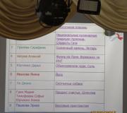
The standard set includes 21 phages (80, 79, 52A, 52, 29, 71, 55, 3C, 3B, 3A, 53,47,42E, 7, 6, 42D, 77.75, 83A, 54, 81, 187.
The inoculated dishes are dried at a temperature of 37° for 30-40 minutes, then a drop of the corresponding phage is applied with a loop, always in the same order.
If there are a lot of cultures, then the cups are placed on the table (in a box) and the lids are removed. Using a Pasteur pipette, take the first and then the next race of test phage and apply small drops to the corresponding square in each dish. At the same time, you should not touch the agar to avoid transfer of the studied cultures from one plate to another. After the phage droplets have dried, the dishes are placed in an inverted position for 5-6 hours in a thermostat (temperature 37°) and left at room temperature until the morning. The results are recorded with the naked eye and with the help of a magnifying glass, noting the number of the phage that gave lysis at + + and higher, and in brackets the number of the phage that gave lysis at + is noted.
This article, like a biology report for grade 5 about bacteriophage viruses, will help the reader learn basic information about these extracellular life forms. Here we will look at their taxonomic location, structural features and vital functions, how they manifest themselves when interacting with bacteria, etc.
Introduction
Everyone knows that the universal representative of the unit of life on planet Earth is the cell. However, the turn between the nineteenth and twentieth centuries was an era during which a number of diseases were discovered that affected animals, plants and even fungi. Analyzing this phenomenon and taking into account general information about human diseases, scientists realized that there are organisms that may be non-cellular in nature.
Such creatures are extremely small in size, and therefore are able to pass through the smallest filter, without stopping where even the smallest cell would stop. This led to the discovery of viruses.
Total information
Before considering representatives of viruses - bacteriophages - let's get acquainted with general information about this kingdom of the taxonomic hierarchy.

DNA (RNA) belonging to the virus, once inside the carrier cell, begins to interact with heredity so that the cell itself begins the uncontrolled process of synthesizing a specific series of proteins encrypted in the nucleic acid of the pathogen itself. Next, replication occurs, performed directly by the cell itself, and thus the process of assembling a new viral particle begins.
Bacteriophage
What are bacteriophage viruses? This is a special form of life on Earth that selectively penetrates bacterial cells. Reproduction most often occurs inside the host, and the process itself leads to lysis. Considering the structure of viruses using the example of bacteriophages, we can conclude that they consist of shells formed by proteins and have an apparatus for reproducing heredity in the form of one RNA chain or two DNA chains. The total number of bacteriophages approximately corresponds to the entire number of bacterial organisms. These viruses take an active part in the chemical circulation of substances and energy in nature. They cause many manifestations of characteristics in bacteria and microbes developed or developing during evolution.
History of discovery

Bacteriology researcher F. Twort created a description of the infectious disease, which he proposed in an article published in 1915. This disease affected staphylococci and could pass through any filters, and could also be transported from one colony of cells to others.
A microbiologist originally from Canada, F. D. Herelle, discovered bacteriophages in September 1917. Their discovery was made independently of the works of F. Twoorot.
In 1897, N. F. Gamaleya became an observer of the phenomenon of bacterial lysis, which occurred under the influence of the process of grafting an agent.

Meaning
The structure of viruses using the example of a bacteriophage can tell us a lot, especially for interaction with other information that a person has about them. For example, they are presumably the oldest form of viral particles. Quantitative analysis tells us that their population has more than 10 30 particles.
In nature, they can be found in the same places where bacteria live, to which they may be sensitive. Since the organisms in question are determined by their habitat, the preferences of the bacteria they infect, it follows that lysing soil bacteria (phages) will live in the soil. The more microorganisms the substrate contains, the more necessary phages there are.
In fact, each bacteriophage embodies one of the basic elemental units of genetic mobility. Using transduction, they cause the emergence of new genes in the hereditary material of the bacterium. About 10 24 bacterial cells can become infected in a second. This form of answer to the question of which viruses are called bacteriophages openly shows us the methods of distribution of hereditary information that occur between bacterial organisms from a common habitat.
Structural features
Answering the question of what structure a bacteriophage virus has, we can conclude that they can be distinguished in accordance with the chemical structure, the type of nucleic acid (NA), morphological data and the form of interaction with bacterial organisms. The size of such an organism can be several thousand times smaller than the microbial cell itself. A typical representative of phages is formed by a head and a tail. The length of the tail section can be two to four times greater than the diameter of the head, in which, by the way, the genetic potential is located, taking the form of a DNA or RNA chain. There is also an enzyme, transcriptase, immersed in an inactive state and surrounded by a shell of proteins or lipoproteins. It determines the storage of the genome inside the cell and is called the capsid.
The structural features of the bacteriophage virus determine its tail compartment as a tube of proteins, which serves as a continuation of the shell that makes up the head. In the region of the tail base there is an ATPase that regenerates energy resources spent on the process of injection of genetic material.
Systematic data

A bacteriophage is a virus that infects bacteria. This is how taxonomy classifies it in a table of hierarchical order. The assignment of the title to them in this science was due to the discovery of a huge number of these organisms. Currently, these issues are being addressed by ICTV. In accordance with the International Standards for the Classification and Distribution of Taxa Among Viruses, bacteriophages are distinguished by the type of nucleic acid they contain or morphological features.
Today, 20 families can be distinguished, among which only 2 belong to those containing RNA and 5 with the presence of an envelope. Among DNA viruses, only 2 families have a single-stranded genome. 9 (the genome appears to us as a circular molecule of deoxyribonucleic acid) and the other 9 with a linear figure. 9 families are specific to bacteria, and the other 9 are specific to archaea.
Effect on the bacterial cell
Bacteriophage viruses, depending on the nature of their interaction with the bacterial cell, can be classified into virulent and moderate type phages. The former are able to increase their number only with the help of lytic cycles. The processes during which the interaction of a virulent phage and a cell occurs consists of adsorption on the cell surface, penetration into the cellular structure, processes for the biosynthesis of phage elements and their bringing into a functional state, as well as the exit of the bacteriophage beyond the host.
Let us consider the description of bacteriophage viruses, based on their further effects in the cell.
Bacteria have special phage-specific structures on their surface, presented in the form of receptors, to which, in fact, the bacteriophage is attached. Using the tail, the phage, through the enzymes contained at its end, destroys the membrane in a certain location of the cell. Next, it contracts, as a result of which DNA is introduced into the cell. The “body” of the bacteriophage virus with its protein shell remains outside.
The injection made by the phage causes a complete restructuring of all metabolic processes. The synthesis of bacterial proteins, as well as RNA and DNA, is completed, and the bacteriophage itself begins the process of transcription thanks to the activity of a personal enzyme called transcriptase, which is activated only after penetration into the bacterial cell.
Both early and late strands of messenger RNA are synthesized after they enter the ribosome of the carrier cell. There, the process of synthesis of such structures as nuclease, ATPase, lysozyme, capsid, tail extension and even DNA polymerase occurs. The replication process proceeds in accordance with a semi-conservative mechanism and is carried out only in the presence of polymerase. Late proteins are formed after the completion of deoxyribonucleic acid replication processes. After this, the final stage of the cycle begins, in which phage maturation occurs. It can also combine with the protein shell and form mature particles ready for infection.
Cycles of life

Regardless of the structure of the bacteriophage virus, they all have a common characteristic of life cycles. According to moderation or virulence, both types of organisms are similar to each other in the initial stages of influencing the cell with the same cycle:
- the process of phage adsorption on a special receptor;
- injecting nucleic acids into the victim;
- the joint process of replication of nucleic acids, both phage and bacteria, starts;
- process of cell division;
- development by lysogenic or lytic route.
The temperate bacteriophage maintains the prophage mode and follows the lysogenic path. Virulent representatives develop in accordance with the lytic model, in which there are a number of sequential processes:
Bacteriophage viruses are widely used in antibacterial therapy, which serves as an alternative to antibiotics. Among the organisms that may be applicable, the most commonly identified are: streptococcal, staphylococcal, Klebsiella, coli, Proteaceae, pyobacteriophages, polyproteinaceae and dysenteria.

On the territory of the Russian Federation, thirteen phage-based medicinal substances have been registered and used in practice for medical purposes. As a rule, such methods of fighting infections are used in cases where the traditional form of treatment does not lead to significant changes, which is due to the weak sensitivity of the pathogen to the antibiotic itself or complete resistance. In practice, the use of bacteriophages leads to the rapid and high-quality achievement of the desired success, but this requires the presence of a biological membrane covered with a layer of polysaccharides, through which antibiotics cannot penetrate.
The therapeutic type of use of phage representatives is not supported in the West. However, it is often used to combat bacteria that cause food poisoning. Many years of experiments in studying the activity of bacteriophages show us that the presence, for example, in the common space of cities and villages determines the subjection of space to preventive measures.
Genetic engineers use bacteriophages as vectors that transfer sections of DNA. And also with their participation, the transfer of genomic information between interacting bacterial cells occurs.