The structure of a human cell and the functions of its organelles. Structure of eukaryotic cells
We invite you to familiarize yourself with the materials and.
: cellulose membrane, membrane, cytoplasm with organelles, nucleus, vacuoles with cell sap.The presence of plastids is the main feature of a plant cell.
Functions of the cell membrane- determines the shape of the cell, protects against environmental factors.
Plasma membrane- a thin film, consisting of interacting molecules of lipids and proteins, delimits the internal contents from the external environment, ensures the transport of water, minerals and organic substances into the cell by osmosis and active transport, and also removes waste products.
Cytoplasm- the internal semi-liquid environment of the cell, in which the nucleus and organelles are located, provides connections between them, and participates in basic life processes.
Endoplasmic reticulum- a network of branching channels in the cytoplasm. It is involved in the synthesis of proteins, lipids and carbohydrates, and in the transport of substances. Ribosomes are bodies located on the ER or in the cytoplasm, consisting of RNA and protein, and are involved in protein synthesis. EPS and ribosomes are a single apparatus for the synthesis and transport of proteins.
Mitochondria- organelles delimited from the cytoplasm by two membranes. Organic substances are oxidized in them and ATP molecules are synthesized with the participation of enzymes. Increase in the surface of the inner membrane on which enzymes are located due to cristae. ATP is an energy-rich organic substance.
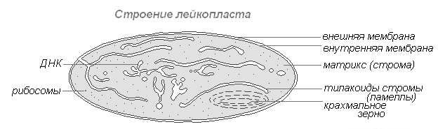
Plastids(chloroplasts, leucoplasts, chromoplasts), their content in the cell is the main feature of the plant organism. Chloroplasts are plastids containing the green pigment chlorophyll, which absorbs light energy and uses it to synthesize organic substances from carbon dioxide and water. Chloroplasts are separated from the cytoplasm by two membranes, numerous outgrowths - grana on the inner membrane, in which chlorophyll molecules and enzymes are located.
Golgi complex- a system of cavities delimited from the cytoplasm by a membrane. The accumulation of proteins, fats and carbohydrates in them. Carrying out the synthesis of fats and carbohydrates on membranes.
Lysosomes- bodies delimited from the cytoplasm by a single membrane. The enzymes they contain accelerate the breakdown of complex molecules into simple ones: proteins into amino acids, complex carbohydrates into simple ones, lipids into glycerol and fatty acids, and also destroy dead parts of the cell and entire cells.
Vacuoles- cavities in the cytoplasm filled with cell sap, a place of accumulation of reserve nutrients and harmful substances; they regulate the water content in the cell.
Core- the main part of the cell, covered on the outside with a two-membrane, pore-pierced nuclear envelope. Substances enter the core and are removed from it through the pores. Chromosomes are carriers of hereditary information about the characteristics of an organism, the main structures of the nucleus, each of which consists of one DNA molecule combined with proteins. The nucleus is the site of DNA, mRNA, and rRNA synthesis.

The presence of an outer membrane, cytoplasm with organelles, and a nucleus with chromosomes.
Outer or plasma membrane- delimits the contents of the cell from the environment (other cells, intercellular substance), consists of lipid and protein molecules, ensures communication between cells, transport of substances into the cell (pinocytosis, phagocytosis) and out of the cell.
Cytoplasm- the internal semi-liquid environment of the cell, which provides communication between the nucleus and organelles located in it. The main life processes take place in the cytoplasm.
Cell organelles:
1) endoplasmic reticulum (ER)- a system of branching tubules, participates in the synthesis of proteins, lipids and carbohydrates, in the transport of substances in the cell;
2) ribosomes- bodies containing rRNA are located on the ER and in the cytoplasm and participate in protein synthesis. EPS and ribosomes are a single apparatus for protein synthesis and transport;
3) mitochondria- “power stations” of the cell, delimited from the cytoplasm by two membranes. The inner one forms cristae (folds), increasing its surface. Enzymes on the cristae accelerate the oxidation of organic substances and the synthesis of energy-rich ATP molecules;
4) Golgi complex- a group of cavities delimited by a membrane from the cytoplasm, filled with proteins, fats and carbohydrates, which are either used in vital processes or removed from the cell. The membranes of the complex carry out the synthesis of fats and carbohydrates;
5) lysosomes- bodies filled with enzymes accelerate the breakdown of proteins into amino acids, lipids into glycerol and fatty acids, polysaccharides into monosaccharides. In lysosomes, dead parts of the cell, whole cells, are destroyed.
Cellular inclusions- accumulations of reserve nutrients: proteins, fats and carbohydrates.
Core- the most important part of the cell. It is covered with a double-membrane shell with pores, through which some substances penetrate into the nucleus, and others enter the cytoplasm. Chromosomes are the main structures of the nucleus, carriers of hereditary information about the characteristics of the organism. It is transmitted during the division of the mother cell to daughter cells, and with germ cells to daughter organisms. The nucleus is the site of DNA, mRNA, and rRNA synthesis.
Exercise:
Explain why organelles are called specialized cell structures?
Answer: organelles are called specialized cell structures, since they perform strictly defined functions, hereditary information is stored in the nucleus, ATP is synthesized in mitochondria, photosynthesis occurs in chloroplasts, etc.
If you have questions about cytology, you can contact
Organoids- permanent, necessarily present, components of the cell that perform specific functions.
Endoplasmic reticulum
Endoplasmic reticulum (ER), or endoplasmic reticulum (ER), is a single-membrane organelle. It is a system of membranes that form “cisterns” and channels, connected to each other and delimiting a single internal space - the EPS cavities. The membranes are connected on one side to the cytoplasmic membrane and on the other to the outer nuclear membrane. There are two types of EPS: 1) rough (granular), containing ribosomes on its surface, and 2) smooth (agranular), the membranes of which do not carry ribosomes.
Functions: 1) transport of substances from one part of the cell to another, 2) division of the cell cytoplasm into compartments (“compartments”), 3) synthesis of carbohydrates and lipids (smooth ER), 4) protein synthesis (rough ER), 5) place of formation of the Golgi apparatus .
Or Golgi complex, is a single-membrane organelle. It consists of stacks of flattened “cisterns” with widened edges. Associated with them is a system of small single-membrane vesicles (Golgi vesicles). Each stack usually consists of 4-6 “cisterns”, is a structural and functional unit of the Golgi apparatus and is called a dictyosome. The number of dictyosomes in a cell ranges from one to several hundred. In plant cells, dictyosomes are isolated.
The Golgi apparatus is usually located near the cell nucleus (in animal cells, often near the cell center).
Functions of the Golgi apparatus: 1) accumulation of proteins, lipids, carbohydrates, 2) modification of incoming organic substances, 3) “packaging” of proteins, lipids, carbohydrates into membrane vesicles, 4) secretion of proteins, lipids, carbohydrates, 5) synthesis of carbohydrates and lipids, 6) place of formation lysosomes The secretory function is the most important, therefore the Golgi apparatus is well developed in secretory cells.
Lysosomes
Lysosomes- single-membrane organelles. They are small bubbles (diameter from 0.2 to 0.8 microns) containing a set of hydrolytic enzymes. Enzymes are synthesized on the rough ER and move to the Golgi apparatus, where they are modified and packaged into membrane vesicles, which, after separation from the Golgi apparatus, become lysosomes themselves. A lysosome can contain from 20 to 60 different types of hydrolytic enzymes. The breakdown of substances using enzymes is called lysis.
There are: 1) primary lysosomes, 2) secondary lysosomes. Primary are called lysosomes that are detached from the Golgi apparatus. Primary lysosomes are a factor ensuring the exocytosis of enzymes from the cell.
Secondary are called lysosomes formed as a result of the fusion of primary lysosomes with endocytic vacuoles. In this case, they digest substances that enter the cell by phagocytosis or pinocytosis, so they can be called digestive vacuoles.
Autophagy- the process of destroying structures unnecessary for the cell. First, the structure to be destroyed is surrounded by a single membrane, then the resulting membrane capsule merges with the primary lysosome, resulting in the formation of a secondary lysosome (autophagic vacuole), in which this structure is digested. The products of digestion are absorbed by the cell cytoplasm, but some of the material remains undigested. The secondary lysosome containing this undigested material is called a residual body. By exocytosis, undigested particles are removed from the cell.
Autolysis- cell self-destruction, which occurs due to the release of lysosome contents. Normally, autolysis occurs during metamorphosis (disappearance of the tail in a tadpole of frogs), involution of the uterus after childbirth, and in areas of tissue necrosis.
Functions of lysosomes: 1) intracellular digestion of organic substances, 2) destruction of unnecessary cellular and non-cellular structures, 3) participation in the processes of cell reorganization.
Vacuoles
Vacuoles- single-membrane organelles are “containers” filled with aqueous solutions of organic and inorganic substances. The ER and Golgi apparatus take part in the formation of vacuoles. Young plant cells contain many small vacuoles, which then, as the cells grow and differentiate, merge with each other and form one large central vacuole. The central vacuole can occupy up to 95% of the volume of a mature cell; the nucleus and organelles are pushed towards the cell membrane. The membrane bounding the plant vacuole is called the tonoplast. The fluid that fills a plant vacuole is called cell sap. The composition of cell sap includes water-soluble organic and inorganic salts, monosaccharides, disaccharides, amino acids, final or toxic metabolic products (glycosides, alkaloids), and some pigments (anthocyanins).
Animal cells contain small digestive and autophagy vacuoles, which belong to the group of secondary lysosomes and contain hydrolytic enzymes. Unicellular animals also have contractile vacuoles that perform the function of osmoregulation and excretion.
Functions of the vacuole: 1) accumulation and storage of water, 2) regulation of water-salt metabolism, 3) maintenance of turgor pressure, 4) accumulation of water-soluble metabolites, reserve nutrients, 5) coloring of flowers and fruits and thereby attracting pollinators and seed dispersers, 6) see. functions of lysosomes.
The endoplasmic reticulum, Golgi apparatus, lysosomes and vacuoles form single vacuolar network of the cell, the individual elements of which can transform into each other.
Mitochondria

1 - outer membrane;
2 - internal membrane; 3 - matrix; 4 - crista; 5 - multienzyme system; 6 - circular DNA.
The shape, size and number of mitochondria vary enormously. Mitochondria can be rod-shaped, round, spiral, cup-shaped, or branched in shape. The length of mitochondria ranges from 1.5 to 10 µm, diameter - from 0.25 to 1.00 µm. The number of mitochondria in a cell can reach several thousand and depends on the metabolic activity of the cell.
The mitochondrion is bounded by two membranes. The outer membrane of mitochondria (1) is smooth, the inner (2) forms numerous folds - cristas(4). Cristae increase the surface area of the inner membrane, on which multienzyme systems (5) involved in the synthesis of ATP molecules are located. The internal space of mitochondria is filled with matrix (3). The matrix contains circular DNA (6), specific mRNA, prokaryotic type ribosomes (70S type), and Krebs cycle enzymes.
Mitochondrial DNA is not associated with proteins (“naked”), is attached to the inner membrane of the mitochondrion and carries information about the structure of about 30 proteins. To build a mitochondrion, many more proteins are required, so information about most mitochondrial proteins is contained in nuclear DNA, and these proteins are synthesized in the cytoplasm of the cell. Mitochondria are capable of autonomous reproduction by fission in two. Between the outer and inner membranes there is proton reservoir, where H + accumulation occurs.
Functions of mitochondria: 1) ATP synthesis, 2) oxygen breakdown of organic substances.
According to one hypothesis (the theory of symbiogenesis), mitochondria originated from ancient free-living aerobic prokaryotic organisms, which, having accidentally penetrated the host cell, then formed a mutually beneficial symbiotic complex with it. The following data support this hypothesis. Firstly, mitochondrial DNA has the same structural features as the DNA of modern bacteria (closed in a ring, not associated with proteins). Secondly, mitochondrial ribosomes and bacterial ribosomes belong to the same type - the 70S type. Thirdly, the mechanism of mitochondrial fission is similar to that of bacteria. Fourth, the synthesis of mitochondrial and bacterial proteins is suppressed by the same antibiotics.
Plastids

1 - outer membrane; 2 - internal membrane; 3 - stroma; 4 - thylakoid; 5 - grana; 6 - lamellae; 7 - starch grains; 8 - lipid drops.
Plastids are characteristic only of plant cells. Distinguish three main types of plastids: leucoplasts are colorless plastids in the cells of uncolored parts of plants, chromoplasts are colored plastids usually yellow, red and orange, chloroplasts are green plastids.
Chloroplasts. In the cells of higher plants, chloroplasts have the shape of a biconvex lens. The length of chloroplasts ranges from 5 to 10 µm, diameter - from 2 to 4 µm. Chloroplasts are bounded by two membranes. The outer membrane (1) is smooth, the inner (2) has a complex folded structure. The smallest fold is called thylakoid(4). A group of thylakoids arranged like a stack of coins is called facet(5). The chloroplast contains on average 40-60 grains, arranged in a checkerboard pattern. The granae are connected to each other by flattened channels - lamellae(6). The thylakoid membranes contain photosynthetic pigments and enzymes that provide ATP synthesis. The main photosynthetic pigment is chlorophyll, which determines the green color of chloroplasts.
The interior space of the chloroplasts is filled stroma(3). The stroma contains circular “naked” DNA, 70S-type ribosomes, Calvin cycle enzymes, and starch grains (7). Inside each thylakoid there is a proton reservoir, and H + accumulates. Chloroplasts, like mitochondria, are capable of autonomous reproduction by dividing into two. They are found in the cells of the green parts of higher plants, especially many chloroplasts in leaves and green fruits. Chloroplasts of lower plants are called chromatophores.
Function of chloroplasts: photosynthesis. It is believed that chloroplasts originated from ancient endosymbiotic cyanobacteria (symbiogenesis theory). The basis for this assumption is the similarity of chloroplasts and modern bacteria in a number of characteristics (circular, “naked” DNA, 70S-type ribosomes, method of reproduction).
Leukoplasts. The shape varies (spherical, round, cupped, etc.). Leukoplasts are bounded by two membranes. The outer membrane is smooth, the inner one forms few thylakoids. The stroma contains circular “naked” DNA, 70S-type ribosomes, enzymes for the synthesis and hydrolysis of reserve nutrients. There are no pigments. The cells of the underground organs of the plant (roots, tubers, rhizomes, etc.) have especially many leucoplasts. Function of leucoplasts: synthesis, accumulation and storage of reserve nutrients. Amyloplasts- leukoplasts that synthesize and accumulate starch, elaioplasts- oils, proteinoplasts- proteins. Different substances can accumulate in the same leukoplast.
Chromoplasts. Bounded by two membranes. The outer membrane is smooth, the inner membrane is either smooth or forms single thylakoids. The stroma contains circular DNA and pigments - carotenoids, which give chromoplasts a yellow, red or orange color. The form of accumulation of pigments is different: in the form of crystals, dissolved in lipid droplets (8), etc. Contained in the cells of mature fruits, petals, autumn leaves, and rarely - root vegetables. Chromoplasts are considered the final stage of plastid development.
Function of chromoplasts: coloring flowers and fruits and thereby attracting pollinators and seed dispersers.
All types of plastids can be formed from proplastids. Proplastids- small organelles contained in meristematic tissues. Since plastids have a common origin, interconversions between them are possible. Leukoplasts can turn into chloroplasts (greening of potato tubers in the light), chloroplasts - into chromoplasts (yellowing of leaves and reddening of fruits). The transformation of chromoplasts into leucoplasts or chloroplasts is considered impossible.
Ribosomes

1 - large subunit; 2 - small subunit.
Ribosomes- non-membrane organelles, diameter approximately 20 nm. Ribosomes consist of two subunits - large and small, into which they can dissociate. The chemical composition of ribosomes is proteins and rRNA. rRNA molecules make up 50-63% of the mass of the ribosome and form its structural framework. There are two types of ribosomes: 1) eukaryotic (with sedimentation constants for the whole ribosome - 80S, small subunit - 40S, large - 60S) and 2) prokaryotic (70S, 30S, 50S, respectively).
Ribosomes of the eukaryotic type contain 4 rRNA molecules and about 100 protein molecules, while the prokaryotic type contains 3 rRNA molecules and about 55 protein molecules. During protein biosynthesis, ribosomes can “work” individually or combine into complexes - polyribosomes (polysomes). In such complexes they are linked to each other by one mRNA molecule. Prokaryotic cells have only 70S-type ribosomes. Eukaryotic cells have both 80S-type ribosomes (rough EPS membranes, cytoplasm) and 70S-type (mitochondria, chloroplasts).
Eukaryotic ribosomal subunits are formed in the nucleolus. The combination of subunits into a whole ribosome occurs in the cytoplasm, usually during protein biosynthesis.
Function of ribosomes: assembly of a polypeptide chain (protein synthesis).
Cytoskeleton
Cytoskeleton formed by microtubules and microfilaments. Microtubules are cylindrical, unbranched structures. The length of microtubules ranges from 100 µm to 1 mm, the diameter is approximately 24 nm, and the wall thickness is 5 nm. The main chemical component is the protein tubulin. Microtubules are destroyed by colchicine. Microfilaments are filaments with a diameter of 5-7 nm and consist of the protein actin. Microtubules and microfilaments form complex weaves in the cytoplasm. Functions of the cytoskeleton: 1) determination of the shape of the cell, 2) support for organelles, 3) formation of the spindle, 4) participation in cell movements, 5) organization of cytoplasmic flow.

Includes two centrioles and a centrosphere. Centriole is a cylinder, the wall of which is formed by nine groups of three fused microtubules (9 triplets), interconnected at certain intervals by cross-links. Centrioles are united in pairs where they are located at right angles to each other. Before cell division, centrioles diverge to opposite poles, and a daughter centriole appears near each of them. They form a division spindle, which contributes to the even distribution of genetic material between daughter cells. In the cells of higher plants (gymnosperms, angiosperms), the cell center does not have centrioles. Centrioles are self-replicating organelles of the cytoplasm; they arise as a result of duplication of existing centrioles. Functions: 1) ensuring the divergence of chromosomes to the cell poles during mitosis or meiosis, 2) the center of organization of the cytoskeleton.
Organoids of movement
Not present in all cells. Organelles of movement include cilia (ciliates, epithelium of the respiratory tract), flagella (flagellates, sperm), pseudopods (rhizopods, leukocytes), myofibrils (muscle cells), etc.
Flagella and cilia- filament-shaped organelles, representing an axoneme bounded by a membrane. Axoneme is a cylindrical structure; the wall of the cylinder is formed by nine pairs of microtubules; in its center there are two single microtubules. At the base of the axoneme there are basal bodies, represented by two mutually perpendicular centrioles (each basal body consists of nine triplets of microtubules; there are no microtubules in its center). The length of the flagellum reaches 150 microns, the cilia are several times shorter.
Myofibrils consist of actin and myosin myofilaments that provide contraction of muscle cells.
Go to lectures No. 6“Eukaryotic cell: cytoplasm, cell membrane, structure and functions of cell membranes”
Cell– an elementary unit of a living system. Specific functions in a cell are distributed between organoids– intracellular structures. Despite the variety of forms, cells of different types have striking similarities in their main structural features.
Cell theory
As microscopes improved, new information appeared about the cellular structure of plant and animal organisms.
With the advent of physical and chemical research methods in cell science, an amazing unity in the structure of cells of different organisms was revealed, and the inextricable connection between their structure and function was proven.
Basic principles of cell theory
The cell is the basic unit of structure and development of all living organisms. The cells of all single- and multicellular organisms are similar in their structure, chemical composition, basic manifestations of life activity and metabolism. Cells reproduce by division. In multicellular organisms, cells are specialized in their functions and form tissues. Organs are made up of tissues.
To confirm some of the above provisions of the cell theory, let us name the common features characteristic of animal and plant cells.
Common characteristics of plant and animal cells
Unity of structural systems - cytoplasm and nucleus. The similarity of metabolic and energy processes. Unity of the principle of hereditary code. Universal membrane structure. Unity of chemical composition. Similarities in the process of cell division.
Table Distinctive features of plant and animal cells
Signs | plant cell | animal cell |
Plastids | Chloroplasts, chromoplasts, leucoplasts | Absent |
Nutrition method | Autotrophic (phototrophic, chemotrophic). | Heterotrophic (saprotrophic, chemotrophic). |
ATP synthesis | In chloroplasts, mitochondria. | In mitochondria. |
ATP breakdown | In chloroplasts and all parts of the cell where energy is required. |
|
Cell center | In lower plants. | In all cells. |
Cellulose cell wall | Located outside the cell membrane. | Absent. |
Inclusion | Spare nutrients in the form of grains of starch, protein, drops of oil; in vacuoles with cell sap; salt crystals. | Spare nutrients in the form of grains and drops (proteins, fats, carbohydrate glycogen); end products of metabolism, salt crystals; pigments. |
Large cavities filled with cell sap - an aqueous solution of various substances that are reserve or final products. Osmotic reservoirs of the cell. | Contractile, digestive, excretory vacuoles. Usually small. |
The importance of theory: it proves the unity of origin of all living organisms on Earth.
Cellular structures
Figure Scheme of the structure of animal and plant cells

Organelles | Structure | Functions |
Cytoplasm | Located between the plasma membrane and the nucleus, it includes various organelles. The space between the organelles is filled with cytosol - a viscous aqueous solution of various salts and organic substances, permeated with a system of protein threads - the cytoskeleton. | Most of the chemical and physiological processes of the cell take place in the cytoplasm. Cytoplasm unites all cellular structures into a single system and ensures the relationship between the exchange of substances and energy between the organelles of the cell. |
Outer cell membrane | An ultramicroscopic film consisting of two monomolecular layers of protein and a bimolecular layer of lipids located between them. The integrity of the lipid layer can be interrupted by protein molecules - “pores”. | Isolates the cell from the environment, has selective permeability, regulates the process of substances entering the cell; ensures the exchange of substances and energy with the external environment, promotes the connection of cells in tissue, participates in pinocytosis and phagocytosis; regulates the water balance of the cell and removes waste products from it. |
Endoplasmic reticulum (ER) | Ultramicroscopic system of membranes forming tubes, tubules, cisterns, vesicles. The structure of the membranes is universal (as well as the outer one), the entire network is united into a single whole with the outer membrane of the nuclear membrane and the outer cellular membrane. The granular ES carries ribosomes, while the smooth one lacks them. | Provides transport of substances both within the cell and between neighboring cells. Divides the cell into separate sections in which various physiological processes and chemical reactions occur simultaneously. Granular ES is involved in protein synthesis. Complex protein molecules are formed in ES channels, fats are synthesized, and ATP is transported. |
Ribosomes | Small spherical organelles consisting of rRNA and protein. | Proteins are synthesized on ribosomes. |
Golgi apparatus | Microscopic single-membrane organelles, consisting of a stack of flat cisterns, along the edges of which tubes branch off, separating small vesicles. | In the general system of membranes of any cells, it is the most mobile and changing organelle. The cisterns accumulate decomposition synthesis products and substances that enter the cell, as well as substances that are removed from the cell. Packed in vesicles, they enter the cytoplasm: some are used, while others are excreted. |
Lysosomes | Microscopic single-membrane organelles of round shape. Their number depends on the vital activity of the cell and its physiological state. Lysosomes contain lysing (dissolving) enzymes synthesized on ribosomes. | Digestion of food that enters an animal cell during phagocytosis and pinocytosis. Protective function. In the cells of any organisms, autolysis (self-dissolution of organelles) occurs; especially under conditions of food or oxygen starvation, the tail of animals dissolves. In plants, organelles dissolve during the formation of cork tissue of wood vessels. |
Conclusions from the lecture
An important achievement of biological science is the formation of ideas about the structure and vital activity of the cell as a structural and functional unit of the body. The science that studies the living cell in all its manifestations is called cytology. The first stages of the development of cytology as a field of scientific knowledge were associated with the works of R. Hooke, A. Leeuwenhoek, T. Schwann, M. Schleiden, R. Virchow, K. Baer. The result of their activity was the formulation and development of the basic principles of cell theory. A variety of cellular structures are directly involved in the vital processes of a cell. Cytoplasm ensures the activity of all cellular structures as a single system. The cytoplasmic membrane ensures the passage selectivity of substances in the cell and protects it from the external environment. The ES ensures the transport of substances both within the cell and between neighboring cells. The products of synthesis and breakdown of substances entering the cell, as well as substances that are removed from the cell, accumulate in the tanks of the Golgi Apparatus. Lysosomes break down substances that enter the cell.
Questions for self-control
Using knowledge of cell theory, prove the unity of the origin of life on Earth. What are the similarities and differences in the structure of plant and animal cells? How is the structure of the cell membrane related to its functions? How does active absorption of substances into cells occur? What is the connection between ribosomes and ES? What are the structure and functions of lysosomes in a cell?
Cellular structures: mitochondria, plastids, organelles of movement, inclusions. Core
Table Cell organelles, their structure and functions
Organelles | Structure | Functions |
Mitochondria | Microscopic organelles with a double-membrane structure. The outer membrane is smooth, the inner one forms outgrowths of various shapes - cristae. The mitochondrial matrix (a semi-liquid substance) contains enzymes, ribosomes, DNA, and RNA. | The universal organelle is a respiratory and energy center. During the oxygen (oxidative) stage in the matrix, with the help of enzymes, organic substances are broken down with the release of energy, which goes to the synthesis of ATP on (cristae). |
Leukoplasts | Microscopic organelles with a double-membrane structure. The inner membrane forms 2–3 outgrowths. The shape is round. Colorless. | Characteristic of plant cells. They serve as a site for the deposition of reserve nutrients, mainly starch grains. In the light, their structure becomes more complex, and they transform into chloroplasts. Formed from proplastids. |
Chloroplasts | Microscopic organelles with a double-membrane structure. The outer membrane is smooth. The inner membrane forms a system of two-layer plates - stromal thylakoids and granal thylakoids. Pigments - chlorophyll and carotenoids - are concentrated in the membranes of thylakoid granules between layers of protein and lipid molecules. The protein-lipid matrix contains its own ribosomes, DNA, and RNA. | Characteristic of plant cells are photosynthesis organelles that are capable of creating organic substances - carbohydrates and free oxygen - from inorganic substances (CO2 and H2O) in the presence of light energy and the pigment chlorophyll. Synthesis of own proteins. They can be formed from plastids or leucoplasts, and in the fall they turn into chloroplasts (red and orange fruits, red and yellow leaves). |
Chromoplasts | Microscopic organelles with a double-membrane structure. Chromoplasts themselves have a spherical shape, and those formed from chloroplasts take the form of caratinodon crystals, typical for this type of plant. Color: red, orange, yellow. | Characteristic of plant cells. They give flower petals a color that is attractive to pollinating insects. Autumn leaves and ripe fruits separated from plants contain crystalline carotenoids - end products of metabolism. |
Cell center | Ultramicroscopic organelle of non-membrane structure. Consists of two centrioles. Each has a cylindrical shape, the walls are formed by nine triplets of tubes, and in the middle there is a homogeneous substance. The centrioles are located perpendicular to each other. | Takes part in the division of cells of animals and lower plants. At the beginning of division (in prophase), the centrioles diverge to different poles of the cell. The spindle strands extend from the centrioles to the centromeres of the chromosomes. In anaphase, these threads attract chromatids to the poles. After the end of division, the centrioles remain in the daughter cells. They double and form a cell center. |
Cellular inclusions (non-permanent structures) | Dense, granular inclusions with a membrane (for example, vacuoles). | |
Organoids of movement | Cilia are numerous cytoplasmic projections on the surface of the membrane. | Removal of dust particles (ciliated epithelium of the upper respiratory tract), movement (single-celled organisms). |
Flagella are single cytoplasmic projections on the cell surface. | Movement (spermatozoa, zoospores, single-celled organisms). |
|
False legs (pseudopodia) are amoeboid protrusions of the cytoplasm. | They are formed in animals in different places of the cytoplasm to capture food and for movement. |
|
Myofibrils are thin filaments up to 1 cm long or more. | They serve to contract the muscle fibers along which they are located. |
|
Cytoplasm, which carries out stream and circular movement. | Movement of cell organelles in relation to (during photosynthesis), heat, chemical irritant. |
Figure Scheme of composition and functions of cellular inclusions

Phagocytosis– capture of solid particles by the plasma membrane and draw them inward.
The plasma membrane forms an invagination in the form of a thin tubule into which liquid with substances dissolved in it enters. This method is called pinocenosis.
Core
All organisms that have a cellular structure without a formed nucleus are called prokaryotes. All organisms that have a cellular structure with a nucleus are called eukaryotes.
Table Nuclear structures, their structure and functions
Structures | Structure | Functions |
Nuclear envelope | Double-layer porous. The outer membrane passes into the ES membranes. It is characteristic of all animal and plant cells, except bacteria and blue-green ones, which do not have a nucleus. | Separates the nucleus from the cytoplasm. Regulates the transport of substances from the nucleus to the cytoplasm (RNA and ribosomal subunits) and from the cytoplasm to the nucleus (proteins, fat, carbohydrates, ATP, water, ions). |
Chromosomes (chromatin) | In an interphase cell, chromatin has the form of fine-grained thread-like structures consisting of DNA molecules and a protein sheath. In dividing cells, chromatin structures spiral and form chromosomes. A chromosome consists of two chromatids, and after nuclear division it becomes single chromatid. By the beginning of the next division, a second chromatid is completed on each chromosome. Chromosomes have a primary constriction on which the centromere is located; the constriction divides the chromosome into two arms of equal or different lengths. Nucleolar chromosomes have a secondary constriction. | Chromatin structures are carriers of DNA. DNA consists of sections - genes that carry hereditary information and are transmitted from ancestors to descendants through germ cells. The totality of chromosomes, and, consequently, the genes of the germ cells of the parents, is transmitted to children, which ensures the stability of the characteristics characteristic of a given population or species. DNA and RNA are synthesized in chromosomes, which serves as a necessary factor in the transmission of hereditary information during cell division and the construction of protein molecules. |
A spherical body resembling a ball of thread. Consists of protein and RNA. Formed on the secondary constriction of the nucleolar chromosome. It breaks down when cells divide. | Formation of ribosome halves from rRNA and protein. The halves (subunits) of ribosomes enter the cytoplasm through pores in the nuclear envelope and combine to form ribosomes. |
|
Nuclear juice (karyolymph) | A semi-liquid substance representing a colloidal solution of proteins, nucleic acids, carbohydrates, and mineral salts. The reaction is sour. | Participates in the transport of substances and nuclear structures, fills the space between nuclear structures; During cell division it mixes with the cytoplasm. |
Figure Scheme of the structure of the cell nucleus
Functions of the cell nucleus:
- regulation of metabolic processes in the cell; storage of hereditary information and its reproduction; RNA synthesis; ribosome assembly.
Conclusions from the lecture
In mitochondria, organic substances are broken down and energy is released, which is used for the synthesis of ATP. Plastids play an important role in ensuring the vital processes of the plant cell. Organelles of movement include cellular structures: cilia, flagella, myofibrils. All cellular organisms are divided into prokaryotes (without a nucleus) and eukaryotes (with a nucleus). The nucleus is a structural and functional center that coordinates its metabolism, directing the processes of self-reproduction and storage of hereditary information.
Questions for self-control
Why are mitochondria figuratively called the “power stations” of the cell? What cell structures contribute to its movement? What are cellular inclusions? What is their role? What are the functions of the nucleus in a cell?
Organic substances in the cell (carbohydrates, proteins, lipids, nucleic acids, ATP, vitamins, etc.)
Biological polymers– organic compounds that make up the cells of living organisms. Polymer is a multi-link chain of simple substances – monomers (n ÷ 10 thousand – 100 thousand monomers)

The properties of biopolymers depend on the structure of their molecules, on the number and variety of monomer units.
If the monomers are different, then their repeated alternations in the chain create a regular polymer.
…A – A – B – A – A – B... regular
…A – A – B – B – A – B – A... irregular
Carbohydrates
General formula Сn(H2O)m
Carbohydrates play the role of energy substances in the human body. The most important of them are - sucrose, glucose, fructose, and starch. They are quickly absorbed ("burned") in the body. The exception is cellulose(cellulose), which is especially abundant in plant foods. It is practically not absorbed by the body, but is of great importance: it acts as ballast and helps digestion, mechanically cleansing the mucous membranes of the stomach and intestines. There are a lot of carbohydrates in potatoes and vegetables, cereals, pasta, fruits and bread.
Glucose, ribose, fructose, deoxyribose - monosaccharides
Sucrose - disaccharides
Starch, glycogen, cellulose - polysaccharides
Finding in nature: in plants, fruits, pollen, vegetables (garlic, beets), potatoes, rice, corn, wheat grain, wood...
Their functions:
- energy: oxidation to CO2 and H2O releases energy; excess energy is stored in liver and muscle cells in the form of glycogen; construction: in a plant cell - a strong base of cell walls (cellulose); structural: part of the intercellular substance of the skin, cartilage tendons; recognition by other cells: as part of cell membranes, if separated liver cells are mixed with kidney cells, they will independently separate into two groups due to the interaction of cells of the same type.
Lipids (lipoids, fats)
Lipids include various fats, fat-like substances, phospholipids... All of them are insoluble in water, but soluble in chloroform, ether...
Finding in nature: in animal and human cells in the cell membrane; between the cells is the subcutaneous layer of fat.
Functions:
- thermal insulation (in whales, pinnipeds...); storage nutrient; energy: energy is released during the hydrolysis of fats; structural: some lipids are integral parts of cell membranes.
Fats also serve as a source of energy for the human body. The body stores them “in reserve” and they serve as a long-term energy source. In addition, fats have low thermal conductivity and protect the body from hypothermia. It is not surprising that the traditional diet of northern peoples contains so much animal fat. For people engaged in heavy physical labor, it is also easiest (although not always healthier) to compensate for the energy expended with fatty foods. Fats are part of cell walls, intracellular formations, and nervous tissue. Another function of fats is to supply fat-soluble vitamins and other biologically active substances to the body tissues.
Squirrels
Figure 1.2.1. Protein molecule


If in R we replace one more H with the amino group NH2, we get the amino acid:

Proteins are biopolymers whose monomers are amino acids.
The formation of linear protein molecules occurs as a result of reactions of amino acids with each other.

Sources of proteins can be not only animal products (meat, fish, eggs, cottage cheese), but also plant products, for example, legumes (beans, peas, soybeans, peanuts, which contain up to 22–23% proteins by weight), nuts and mushrooms . However, the most protein is in cheese (up to 25%), meat products (pork 8–15%, lamb 16–17%, beef 16–20%), poultry (21%), fish (13–21%), eggs (13%), cottage cheese (14%). Milk contains 3% proteins, and bread 7–8%. Among cereals, the champion in proteins is buckwheat (13% of proteins in dry cereals), which is why it is recommended for dietary nutrition. To avoid “excesses” and at the same time ensure the normal functioning of the body, it is necessary, first of all, to give a person a complete set of proteins with food. If there is not enough protein in the diet, an adult feels a loss of strength, his performance decreases, and his body is less resistant to infections and colds. As for children, if they have inadequate protein nutrition, they are greatly behind in development: children grow, and proteins are the main “building material” of nature. Every cell of a living organism contains proteins. Human muscles, skin, hair, and nails consist mainly of proteins. Moreover, proteins are the basis of life; they participate in metabolism and ensure the reproduction of living organisms.
Structure:
- primary structure – linear, with alternating amino acids; secondary - in the form of a spiral with weak bonds between the turns (hydrogen); tertiary - a spiral rolled into a ball; quaternary - when combining several chains that differ in primary structure.
With radiation, high temperatures, extreme pH values, in alcohol, acetone, the protein is destroyed - a denaturation reaction.
Table 1.2.1. Protein structure
| Primary structure– a specific sequence of α-amino acid residues in a polypeptide chain |
Secondary structure– conformation of the polypeptide chain, secured by many hydrogen bonds between N-H and C=O groups. One of the models of secondary structure is an α-helix due to cooperative intramolecular H-bonds. Another model is the b-form (“folded sheet”), in which interchain (intermolecular) H-bonds predominate |
|
| Tertiary structure- the shape of a twisted helix in space, formed mainly due to disulfide bridges - S-S-, hydrogen bonds, hydrophobic and ionic interactions |
| Quaternary structure– aggregates of several protein macromolecules (protein complexes), formed through the interaction of different polypeptide chains |


Functions:
- construction: proteins are an essential component of all cellular structures; structural: proteins in combination with DNA make up the body of chromosomes, and with RNA – the body of ribosomes; enzymatic: chemical catalyst. reactions are performed by any enzyme - a protein, but a very specific one; transport: transfer of O2, hormones in the body of animals and humans; regulatory: proteins can perform a regulatory function if they are hormones. For example, insulin (a hormone that supports the functioning of the pancreas) activates the uptake of glucose molecules by cells and their breakdown or storage inside the cell. With a lack of insulin, glucose accumulates in the blood, developing diabetes; protective: when foreign bodies enter the body, protective proteins are produced - antibodies, which bind to foreign bodies, combine and suppress their vital functions. This mechanism of resistance of the body is called immunity; energy: with a lack of carbohydrates and fats, amino acid molecules can be oxidized.
Adenosine triphosphoric acid (ATP)– a universal carrier and main energy accumulator in living maples, which is necessary for the synthesis of organic substances, movement, production of heat, nerve impulses, and luminescence. ATP is found in all plant and animal cells.
It is a nucleotide formed by residues of a nitrogenous base (adenine), a sugar (ribose) and three phosphoric acid residues.
ATP is an unstable molecule: when the terminal phosphoric acid residue is removed. ATP is converted into ADP (adenosine diphosphoric acid), and about 30.5 kJ is released.
Figure 1.2.2. The structure of the ATP molecule

Hormones organic compounds, which may be of a protein nature (pancreatic hormones) and may be lipids (sex hormones), may be derivatives of amino acids. Hormones are produced by both animals and plants. Hormones perform various functions:
- regulate the content of sodium ions and water in the body; ensure puberty; anxiety and stress hormones increase the release of glucose into the blood and, therefore, determine the active use of energy; signaling hormones report the presence of food and danger; Plants have their own hormones that accelerate the ripening of fruits and attract insects.
Nucleic acids– biopolymers whose monomers are nucleotides.
Figure 1.2.3. Nucleic acid synthesis

Figure 1.2.4. Schematic structure of DNA (ellipses indicate hydrogen bonds)

The DNA molecule is a structure consisting of two strands, which are connected to each other along their entire length by hydrogen bonds. (Fig. 1.2.4)
Figure 1.2.5. Section of a DNA molecule

A feature of the DNA structure is that opposite the nitrogenous base A in one chain lies the nitrogenous base T in the other chain, and opposite the nitrogenous base G is always the nitrogenous base C. The above can be shown in the form of a diagram:

These base pairs are called complementary bases (complementary to each other). DNA strands in which the bases are located complementary to each other are called complementary strands. In Fig. Figure 1.2.5 shows two strands of DNA that are connected by complementary regions.
The order of nucleotides in DNA molecules determines the order of amino acids in linear protein molecules.
Table Comparative characteristics of DNA and RNA
Signs of comparison | ||
Location in the cage | Nucleus, mitochondria, chloroplasts | Nucleus, ribosomes, cytoplasm, mitochondria, chloroplasts |
Location in the nucleus | Chromosomes | |
Structure of a macromolecule | Double unbranched linear polymer, coiled in a right-handed helix | Single polynucleotide chain |
Composition of nucotides | Nitrogen base (adenine, guanine, thymine, cytosine); deoxyribose (carbohydrate); phosphoric acid residue | Nitrogen base (adenine, guanine, uracil, cytosine); ribose (carbohydrate); phosphoric acid residue |
Chemical basis of chromosomal genetic material (gene); DNA and RNA synthesis, information about protein structure | Information (mRNA) transmits the code of hereditary information about the primary structure of the protein molecule; ribosomal (rRNA) is part of ribosomes; transport (tRNA) carries amino acids to ribosomes. |
Vitamins
Back in the late 19th century, scientists discovered that the terrible beri-beri disease, which damages the nervous system, is caused by a lack of some special substance in food. In 1912, Polish researcher Kazimierz Funk (1884–1967) isolated a substance from rice bran and called it vitamin (from the Latin vita - “life”). This is the name for chemical compounds that are required for the normal functioning of the body in very small quantities. The body “does not know how” to synthesize vitamins on its own. Therefore, it is very important to replenish the body with vitamin-containing foods. Lack of vitamins in the body is the cause of a serious disease - vitamin deficiency.
A healthy person under normal living conditions should try to fully cover his need for vitamins through a varied and nutritious diet. You should turn to pharmaceutical preparations containing vitamins in cases where you experience a permanent or seasonal (autumn, spring) deficiency of vitamins, as well as under severe stress. Unsystematic amateur “eating” of vitamin pills can cause unpleasant consequences in the form of hypervitaminosis, when even the required amount of vitamins is not absorbed, but is excreted by the body.
Vitamins
Back in the late 19th century, scientists discovered that the terrible beriberi disease, which damages the nervous system, is caused by a lack of some special substance in food. In 1912, Polish researcher Kazimierz Funk (1884–1967) isolated such a substance from rice bran and called it a vitamin (from the Latin vita - “life”). About 25 vitamins have now been well studied. Their chemical composition and names are very complex, so they were assigned alphabetic symbols. It is customary to divide all vitamins into two large groups: water-soluble And fat-soluble.
The main water-soluble vitamins are:
1. B1 – thiamine, first found in white cabbage; then it was also found in some cereals, raw fish, yeast and sprouted wheat. This vitamin regulates metabolism, nervous activity and is responsible for the condition of the cardiovascular system. The lack of B1 in food causes beriberi, a severe joint disease associated with damage to the nervous system, heart and blood vessels. Beriberi is common in those regions of Southeast Asia where the population eats a poor and monotonous diet, mainly only refined rice, which contains almost no vitamin B1. The body's daily need for vitamin B1 is 1.5–2.0 mg.
2. B2 – riboflavin. Regulates metabolism, increases visual acuity, improves liver and nervous system function, as well as skin condition. Sources of vitamin B2 are yeast, meat, fish, liver and other offal (kidneys, heart, tongue), egg yolk, dairy products, legumes and many cereals. The body's daily need for vitamin B2 is 2.0–2.5 mg;
3. RR – a nicotinic acid(niacin) regulates cellular respiration and cardiac activity. Sources of vitamin PP include yeast, meat and dairy products, and grain crops. In addition, it is one of the few vitamins that can be produced in the human body. Vitamin PP is formed from tryptophan, an amino acid that is part of proteins supplied with food. The body's daily need for vitamin PP is 15–20 mg;
4. B6 – pyridoxine, participates in metabolic processes, is necessary for the absorption of amino acids and for the synthesis of vitamin PP from tryptophan. The body's daily need for vitamin B6 is 2 mg;
5. BC – folacin, folic acid and its derivatives, regulate hematopoiesis and fat metabolism. Contained in liver, yeast, and many vegetables (parsley, spinach, and lettuce). The body's daily need for vitamin BC is 2.0–2.5 mg.
6. B12 – cyanocobalamin. Prevents anemia. Present in beef and pork liver, rabbit and chicken meat, eggs, fish, milk. The body's daily requirement for vitamin B12 is 3 mg.
7. C – ascorbic acid, protects against scurvy, improves immunity. Sources of this vitamin in the diet are fresh and canned vegetables, fruits, and berries. Rose hips, currants, parsley, dill are especially rich in ascorbic acid, and among the wild ones there are nettles, sorrel, and wild garlic. Ascorbic acid is unstable: in air it easily oxidizes to dehydroascorbic acid, which does not have vitamin properties. This must be taken into account when cooking vegetables and fruits. The body's daily need for vitamin C is 75–100 mg.
8. R – routine(bioflavonoid) is a vascular strengthening agent, is active together with vitamin C. There is especially a lot of it in currants, rose hips, chokeberry (chokeberry), citrus fruits and green tea. The body's daily need for vitamin P is 25–50 mg.
Among the fat-soluble vitamins, the most important are:
1. A – retinol and its derivatives, improves the condition of the skin and mucous membranes of the eyes, increases immunity, and most importantly, ensures visual acuity in the twilight. With a lack of vitamin A, “night blindness” occurs (a person has difficulty seeing in the evening). Retinol is found in milk, butter, cheese, fish oil, and can also be synthesized in the human liver from provitamin A - carotene, the source of which is carrots, tomatoes and sea buckthorn. The body's daily need for vitamin A is 1.5 - 2.0 mg (or 6 mg of carotene);
2. D – ergocalciferol, has an antirachitic effect and helps the absorption of calcium. It is absolutely necessary for a growing body during the formation and development of bones and teeth. Vitamin D is found in fish oil, caviar, butter, eggs, and milk. In addition, it is formed in the body under the influence of sunlight. The body's daily requirement for vitamin D is 0.01 mg.
3. E – tocopherol, affects the functions of the gonads and promotes the normal course of pregnancy, promotes the absorption of fat-soluble vitamins, and participates in metabolism. Contained in vegetable oil, buckwheat, legumes. The body's daily requirement for vitamin E is 12–15 mg.
4. K – antihemorrhagic factor, regulates blood clotting, prevents bleeding. Sources of this vitamin include potatoes, cabbage, pumpkin, spinach, sorrel, and liver. The body's daily requirement for vitamin K is 0.2–0.3 mg.
Conclusions from the lecture
The main organic substances in the cell include proteins, carbohydrates, fats, nucleic acids and ATP. Carbohydrates play the role of energy substances in the life of plants, animals, fungi and microorganisms. Fats are the main structural component of cell membranes and a source of energy. They undergo complex transformations in the cell. Proteins are biological polymers, the monomers of which are 20 essential amino acids, and perform a number of important functions in the cell. Construction: proteins are an essential component of all cellular structures; structural: proteins in combination with DNA make up the body of chromosomes, and with RNA – the body of ribosomes; enzymatic: chemical catalyst. reactions – specific enzyme – protein; transport: transfer of O2, hormones in the body of animals and humans; regulatory: (hormones) part of hormones - proteins, for example insulin - a hormone that supports glands, activates the uptake of glucose molecules by cells and their breakdown or storage inside the cell. With a lack of insulin, glucose accumulates in the blood, developing diabetes; protective: when foreign bodies enter the body, protective proteins are produced - antibodies, which bind to foreign bodies, combine and suppress their vital activity. This mechanism of resistance of the body is called immunity; energy: with a lack of carbohydrates and fats, amino acid molecules can oxidize. DNA - molecules of heredity, consist of monomers - nucleotides. DNA and RNA nucleotides have similarities and differences in structure and perform different functions. The great importance of vitamins for organisms has been revealed.
Questions for self-control
What carbohydrates are characteristic of a plant cell and an animal cell? Specify the functions of carbohydrates. Describe the structure of protein molecules in connection with their functions in the cell. What is the primary, secondary, tertiary and quaternary structure of a protein molecule? What is special about the structure of the DNA molecule? What components make up nucleotides? What functions do DNA and RNA perform?
Based on materials from the site http://umka. *****
All living beings and organisms do not consist of cells: plants, fungi, bacteria, animals, people. Despite its minimal size, all the functions of the whole organism are performed by the cell. Complex processes take place inside it, on which the vitality of the body and the functioning of its organs depend.
In contact with
Structural features
Scientists are studying structural features of the cell and the principles of its work. A detailed examination of the structural features of a cell is possible only with the help of a powerful microscope.
All our tissues - skin, bones, internal organs consist of cells that are construction material, come in different shapes and sizes, each variety performs a specific function, but the main features of their structure are similar.
First let's find out what's behind it structural organization of cells. In the course of their research, scientists have found that the cellular foundation is membrane principle. It turns out that all cells are formed from membranes, which consist of a double layer of phospholipids, where protein molecules are immersed on the outside and inside.
What property is characteristic of all types of cells: the same structure, as well as functionality - regulation of the metabolic process, use of their own genetic material (presence and RNA), receipt and consumption of energy.
The structural organization of the cell is based on the following elements that perform a specific function:
- membrane- cell membrane, consists of fats and proteins. Its main task is to separate substances inside from the external environment. The structure is semi-permeable: it can also transmit carbon monoxide;
- core– the central region and main component, separated from other elements by a membrane. It is inside the nucleus that there is information about growth and development, genetic material, presented in the form of DNA molecules that make up the composition;
- cytoplasm- this is a liquid substance that forms the internal environment where various vital processes take place and contains many important components.
What does the cellular content consist of, what are the functions of the cytoplasm and its main components:
- Ribosome- the most important organelle that is necessary for the processes of biosynthesis of proteins from amino acids; proteins perform a huge number of vital tasks.
- Mitochondria- another component located inside the cytoplasm. It can be described in one phrase – an energy source. Their function is to provide components with power for further energy production.
- Golgi apparatus consists of 5 - 8 bags that are connected to each other. The main task of this apparatus is to transfer proteins to other parts of the cell to provide energy potential.
- Damaged elements are cleaned lysosomes.
- Handles transportation endoplasmic reticulum, through which proteins move molecules of useful substances.
- Centrioles are responsible for reproduction.
Core
Since it is a cellular center, special attention should be paid to its structure and functions. This component is the most important element for all cells: it contains hereditary characteristics. Without the nucleus, the processes of reproduction and transmission of genetic information would become impossible. Look at the picture depicting the structure of the nucleus.
- The nuclear membrane, which is highlighted in lilac, lets the necessary substances in and releases them back through the pores - small holes.
- Plasma is a viscous substance and contains all other nuclear components.
- the core is located in the very center and has the shape of a sphere. Its main function is the formation of new ribosomes.
- If you examine the central part of the cell in cross-section, you can see subtle blue weaves - chromatin, the main substance, which consists of a complex of proteins and long strands of DNA that carry the necessary information.

Cell membrane
Let's take a closer look at the work, structure and functions of this component. Below is a table that clearly shows the importance of the outer shell.

Chloroplasts
This is another most important component. But why weren’t chloroplasts mentioned earlier, you ask? Yes, because this component is found only in plant cells. The main difference between animals and plants is the method of nutrition: in animals it is heterotrophic, and in plants it is autotrophic. This means that animals are not able to create, that is, synthesize organic substances from inorganic ones - they feed on ready-made organic substances. Plants, on the contrary, are capable of carrying out the process of photosynthesis and contain special components - chloroplasts. These are green plastids containing the substance chlorophyll. With its participation, light energy is converted into the energy of chemical bonds of organic substances.
Interesting! Chloroplasts are concentrated in large quantities mainly in the above-ground parts of plants - green fruits and leaves.
If you are asked the question: name an important feature of the structure of the organic compounds of a cell, then the answer can be given as follows.
- many of them contain carbon atoms, which have different chemical and physical properties, and are also capable of combining with each other;
- are carriers, active participants in various processes occurring in organisms, or are their products. This refers to hormones, various enzymes, vitamins;
- can form chains and rings, which provides a variety of connections;
- are destroyed when heated and interacting with oxygen;
- atoms within molecules are combined with each other using covalent bonds, do not decompose into ions and therefore interact slowly, reactions between substances take a very long time - several hours and even days.

Structure of chloroplast
Fabrics
Cells can exist one at a time, as in unicellular organisms, but most often they combine into groups of their own kind and form various tissue structures that make up the organism. There are several types of tissues in the human body:
- epithelial– concentrated on the surface of the skin, organs, elements of the digestive tract and respiratory system;
- muscular— we move thanks to the contraction of the muscles of our body, we carry out a variety of movements: from the simplest movement of the little finger to high-speed running. By the way, the heartbeat also occurs due to the contraction of muscle tissue;
- connective tissue makes up up to 80 percent of the mass of all organs and plays a protective and supporting role;
- nervous- forms nerve fibers. Thanks to it, various impulses pass through the body.

Reproduction process
Throughout the life of an organism, mitosis occurs - this is the name given to the process of division. consisting of four stages:
- Prophase. The cell's two centrioles divide and move in opposite directions. At the same time, the chromosomes form pairs, and the nuclear shell begins to collapse.
- The second stage is called metaphases. The chromosomes are located between the centrioles, and gradually the outer shell of the nucleus completely disappears.
- Anaphase is the third stage, during which the centrioles continue to move in the opposite direction from each other, and individual chromosomes also follow the centrioles and move away from each other. The cytoplasm and the entire cell begin to shrink.
- Telophase– final stage. The cytoplasm contracts until two identical new cells appear. A new membrane is formed around the chromosomes and one pair of centrioles appears in each new cell.
Interesting! Cells in epithelium divide faster than in bone tissue. It all depends on the density of the fabrics and other characteristics. The average lifespan of the main structural units is 10 days.
Cell structure. Cell structure and functions. Cell life.
Conclusion
You learned what the structure of a cell is - the most important component of the body. Billions of cells make up an amazingly wisely organized system that ensures the performance and vital activity of all representatives of the animal and plant world.
At the dawn of the development of life on Earth, all cellular forms were represented by bacteria. They absorbed organic substances dissolved in the primordial ocean through the surface of the body.
Over time, some bacteria have adapted to produce organic substances from inorganic ones. To do this, they used the energy of sunlight. The first ecological system arose in which these organisms were producers. As a result, oxygen released by these organisms appeared in the Earth's atmosphere. With its help, you can get much more energy from the same food, and use the additional energy to complicate the structure of the body: dividing the body into parts.
One of the important achievements of life is the separation of the nucleus and cytoplasm. The nucleus contains hereditary information. A special membrane around the core made it possible to protect against accidental damage. As needed, the cytoplasm receives commands from the nucleus that direct the life and development of the cell.
Organisms in which the nucleus is separated from the cytoplasm have formed the nuclear superkingdom (these include plants, fungi, and animals).
Thus, the cell - the basis of the organization of plants and animals - arose and developed in the course of biological evolution.
Even with the naked eye, or even better under a magnifying glass, you can see that the flesh of a ripe watermelon consists of very small grains, or grains. These are cells - the smallest “building blocks” that make up the bodies of all living organisms, including plants.
The life of a plant is carried out by the combined activity of its cells, creating a single whole. With multicellularity of plant parts, there is a physiological differentiation of their functions, specialization of various cells depending on their location in the plant body.
A plant cell differs from an animal cell in that it has a dense membrane that covers the internal contents on all sides. The cell is not flat (as it is usually depicted), it most likely looks like a very small bubble filled with mucous contents.
Structure and functions of a plant cell
Let's consider a cell as a structural and functional unit of an organism. The outside of the cell is covered with a dense cell wall, in which there are thinner sections called pores. Beneath it there is a very thin film - a membrane covering the contents of the cell - the cytoplasm. In the cytoplasm there are cavities - vacuoles filled with cell sap. In the center of the cell or near the cell wall there is a dense body - a nucleus with a nucleolus. The nucleus is separated from the cytoplasm by the nuclear envelope. Small bodies called plastids are distributed throughout the cytoplasm.
Structure of a plant cell
Structure and functions of plant cell organelles
| Organoid | Drawing | Description | Function | Peculiarities |
Cell wall or plasma membrane | Colourless, transparent and very durable | Passes substances into and out of the cell. | Cell membrane is semi-permeable |
|
Cytoplasm | Thick viscous substance | All other parts of the cell are located in it | Is in constant motion |
|
Nucleus (important part of the cell) | Round or oval | Ensures the transfer of hereditary properties to daughter cells during division | Central part of the cell |
|
Spherical or irregular in shape | Takes part in protein synthesis | |||
 | A reservoir separated from the cytoplasm by a membrane. Contains cell sap | Spare nutrients and waste products that the cell does not need accumulate. | As the cell grows, small vacuoles merge into one large (central) vacuole |
|
Plastids | Chloroplasts | They use the light energy of the sun and create organic from inorganic | The shape of discs delimited from the cytoplasm by a double membrane |
|
Chromoplasts | Formed as a result of the accumulation of carotenoids | Yellow, orange or brown |
||
 | Leukoplasts | Colorless plastids | ||
Nuclear envelope | Consists of two membranes (outer and inner) with pores | Separates the nucleus from the cytoplasm | Allows exchange between the nucleus and cytoplasm |
The living part of a cell is a membrane-bound, ordered, structured system of biopolymers and internal membrane structures involved in a set of metabolic and energy processes that maintain and reproduce the entire system as a whole.
An important feature is that the cell does not have open membranes with free ends. Cell membranes always limit cavities or areas, closing them on all sides.
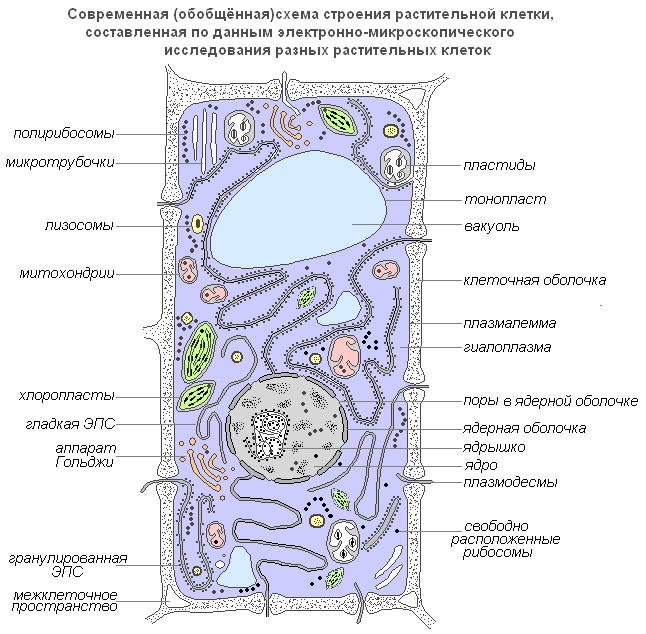
Modern generalized diagram of a plant cell
Plasmalemma(outer cell membrane) is an ultramicroscopic film 7.5 nm thick, consisting of proteins, phospholipids and water. This is a very elastic film that is well wetted by water and quickly restores integrity after damage. It has a universal structure, i.e. typical for all biological membranes. In plant cells, outside the cell membrane there is a strong cell wall that creates external support and maintains the shape of the cell. It consists of fiber (cellulose), a water-insoluble polysaccharide.
Plasmodesmata plant cells, are submicroscopic tubules that penetrate the membranes and are lined with a plasma membrane, which thus passes from one cell to another without interruption. With their help, intercellular circulation of solutions containing organic nutrients occurs. They also transmit biopotentials and other information.
Porami called openings in the secondary membrane, where the cells are separated only by the primary membrane and the median lamina. The areas of the primary membrane and the middle plate separating the adjacent pores of adjacent cells are called the pore membrane or the closing film of the pore. The closing film of the pore is pierced by plasmodesmal tubules, but a through hole is usually not formed in the pores. Pores facilitate the transport of water and solutes from cell to cell. Pores form in the walls of neighboring cells, usually one opposite the other.
Cell membrane has a well-defined, relatively thick shell of a polysaccharide nature. The shell of a plant cell is a product of the activity of the cytoplasm. The Golgi apparatus and the endoplasmic reticulum take an active part in its formation.

Structure of the cell membrane
The basis of the cytoplasm is its matrix, or hyaloplasm, a complex colorless, optically transparent colloidal system capable of reversible transitions from sol to gel. The most important role of hyaloplasm is to unite all cellular structures into a single system and ensure interaction between them in the processes of cellular metabolism.
Hyaloplasma(or cytoplasmic matrix) constitutes the internal environment of the cell. It consists of water and various biopolymers (proteins, nucleic acids, polysaccharides, lipids), of which the main part consists of proteins of varying chemical and functional specificity. The hyaloplasm also contains amino acids, monosaccharides, nucleotides and other low molecular weight substances.
Biopolymers form a colloidal medium with water, which, depending on conditions, can be dense (in the form of a gel) or more liquid (in the form of a sol), both throughout the cytoplasm and in its individual sections. In the hyaloplasm, various organelles and inclusions are localized and interact with each other and the hyaloplasm environment. Moreover, their location is most often specific to certain types of cells. Through the bilipid membrane, the hyaloplasm interacts with the extracellular environment. Consequently, hyaloplasm is a dynamic environment and plays an important role in the functioning of individual organelles and the life of cells in general.
Cytoplasmic formations - organelles
Organelles (organelles) are structural components of the cytoplasm. They have a certain shape and size and are obligatory cytoplasmic structures of the cell. If they are absent or damaged, the cell usually loses its ability to continue to exist. Many of the organelles are capable of division and self-reproduction. Their sizes are so small that they can only be seen with an electron microscope.
Core
The nucleus is the most prominent and usually the largest organelle of the cell. It was first explored in detail by Robert Brown in 1831. The nucleus provides the most important metabolic and genetic functions of the cell. It is quite variable in shape: it can be spherical, oval, lobed, or lens-shaped.
The nucleus plays a significant role in the life of the cell. A cell from which the nucleus has been removed no longer secretes a membrane and stops growing and synthesizing substances. The products of decay and destruction intensify in it, as a result of which it quickly dies. The formation of a new nucleus from the cytoplasm does not occur. New nuclei are formed only by dividing or crushing the old one.
The internal contents of the nucleus are karyolymph (nuclear juice), which fills the space between the structures of the nucleus. It contains one or more nucleoli, as well as a significant number of DNA molecules connected to specific proteins - histones.

Core structure
Nucleolus
The nucleolus, like the cytoplasm, contains predominantly RNA and specific proteins. Its most important function is that it forms ribosomes, which carry out the synthesis of proteins in the cell.
Golgi apparatus
The Golgi apparatus is an organelle that is universally distributed in all types of eukaryotic cells. It is a multi-tiered system of flat membrane sacs, which thicken along the periphery and form vesicular processes. It is most often located near the nucleus.

Golgi apparatus
The Golgi apparatus necessarily includes a system of small vesicles (vesicles), which are detached from thickened cisterns (discs) and are located along the periphery of this structure. These vesicles play the role of an intracellular transport system for specific sector granules and can serve as a source of cellular lysosomes.
The functions of the Golgi apparatus also consist of the accumulation, separation and release outside the cell with the help of vesicles of intracellular synthesis products, decay products, and toxic substances. Products of the cell's synthetic activity, as well as various substances entering the cell from the environment through the channels of the endoplasmic reticulum, are transported to the Golgi apparatus, accumulate in this organelle, and then in the form of droplets or grains enter the cytoplasm and are either used by the cell itself or excreted outside. . In plant cells, the Golgi apparatus contains enzymes for the synthesis of polysaccharides and the polysaccharide material itself, which is used to build the cell wall. It is believed that it is involved in the formation of vacuoles. The Golgi apparatus was named after the Italian scientist Camillo Golgi, who first discovered it in 1897.
Lysosomes
Lysosomes are small vesicles bounded by a membrane whose main function is to carry out intracellular digestion. The use of the lysosomal apparatus occurs during the germination of a plant seed (hydrolysis of reserve nutrients).

Structure of a lysosome
Microtubules
Microtubules are membranous, supramolecular structures consisting of protein globules arranged in spiral or straight rows. Microtubules perform a predominantly mechanical (motor) function, ensuring the mobility and contractility of cell organelles. Located in the cytoplasm, they give the cell a certain shape and ensure the stability of the spatial arrangement of organelles. Microtubules facilitate the movement of organelles to places determined by the physiological needs of the cell. A significant number of these structures are located in the plasmalemma, near the cell membrane, where they participate in the formation and orientation of cellulose microfibrils of plant cell walls.

Microtubule structure
Vacuole
The vacuole is the most important component of plant cells. It is a kind of cavity (reservoir) in the mass of the cytoplasm, filled with an aqueous solution of mineral salts, amino acids, organic acids, pigments, carbohydrates and separated from the cytoplasm by a vacuolar membrane - the tonoplast.
Cytoplasm fills the entire internal cavity only in the youngest plant cells. As the cell grows, the spatial arrangement of the initially continuous mass of cytoplasm changes significantly: small vacuoles filled with cell sap appear, and the entire mass becomes spongy. With further cell growth, individual vacuoles merge, pushing the layers of cytoplasm to the periphery, as a result of which the formed cell usually contains one large vacuole, and the cytoplasm with all organelles is located near the membrane.
Water-soluble organic and mineral compounds of vacuoles determine the corresponding osmotic properties of living cells. This solution of a certain concentration is a kind of osmotic pump for controlled penetration into the cell and release of water, ions and metabolite molecules from it.
In combination with the cytoplasm layer and its membranes, characterized by semi-permeable properties, the vacuole forms an effective osmotic system. Osmotically determined are such indicators of living plant cells as osmotic potential, suction force and turgor pressure.

Structure of the vacuole
Plastids
Plastids are the largest (after the nucleus) cytoplasmic organelles, inherent only in the cells of plant organisms. They are not found only in mushrooms. Plastids play an important role in metabolism. They are separated from the cytoplasm by a double membrane shell, and some types have a well-developed and ordered system of internal membranes. All plastids are of the same origin.
Chloroplasts- the most common and most functionally important plastids of photoautotrophic organisms that carry out photosynthetic processes, ultimately leading to the formation of organic substances and the release of free oxygen. Chloroplasts of higher plants have a complex internal structure.
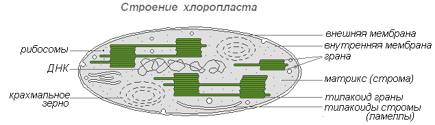
Chloroplast structure
The sizes of chloroplasts in different plants are not the same, but on average their diameter is 4-6 microns. Chloroplasts are able to move under the influence of the movement of the cytoplasm. In addition, under the influence of lighting, active movement of amoeboid-type chloroplasts towards the light source is observed.
Chlorophyll is the main substance of chloroplasts. Thanks to chlorophyll, green plants are able to use light energy.
Leukoplasts(colorless plastids) are clearly defined cytoplasmic bodies. Their sizes are somewhat smaller than the sizes of chloroplasts. Their shape is also more uniform, approaching spherical.
Leukoplast structure
Found in epidermal cells, tubers, and rhizomes. When illuminated, they very quickly turn into chloroplasts with a corresponding change in the internal structure. Leucoplasts contain enzymes with the help of which starch is synthesized from excess glucose formed during photosynthesis, the bulk of which is deposited in storage tissues or organs (tubers, rhizomes, seeds) in the form of starch grains. In some plants, fats are deposited in leucoplasts. The reserve function of leukoplasts occasionally manifests itself in the formation of reserve proteins in the form of crystals or amorphous inclusions.
Chromoplasts in most cases they are derivatives of chloroplasts, occasionally - leucoplasts.

Chromoplast structure
The ripening of rose hips, peppers, and tomatoes is accompanied by the transformation of chloro- or leucoplasts of the pulp cells into caratinoid plasts. The latter contain predominantly yellow plastid pigments - carotenoids, which, when ripe, are intensively synthesized in them, forming colored lipid droplets, solid globules or crystals. In this case, chlorophyll is destroyed.
Mitochondria
Mitochondria are organelles characteristic of most plant cells. They have a variable shape of sticks, grains, and threads. Discovered in 1894 by R. Altman using a light microscope, and the internal structure was studied later using an electron microscope.

The structure of mitochondria
Mitochondria have a double-membrane structure. The outer membrane is smooth, the inner one forms outgrowths of various shapes - tubes in plant cells. The space inside the mitochondrion is filled with semi-liquid content (matrix), which includes enzymes, proteins, lipids, calcium and magnesium salts, vitamins, as well as RNA, DNA and ribosomes. The enzymatic complex of mitochondria accelerates the complex and interconnected mechanism of biochemical reactions that result in the formation of ATP. In these organelles, cells are provided with energy - the energy of chemical bonds of nutrients is converted into high-energy bonds of ATP in the process of cellular respiration. It is in mitochondria that the enzymatic breakdown of carbohydrates, fatty acids, and amino acids occurs with the release of energy and its subsequent conversion into ATP energy. The accumulated energy is spent on growth processes, on new syntheses, etc. Mitochondria multiply by division and live for about 10 days, after which they are destroyed.
Endoplasmic reticulum
The endoplasmic reticulum is a network of channels, tubes, vesicles, and cisterns located inside the cytoplasm. Discovered in 1945 by the English scientist K. Porter, it is a system of membranes with an ultramicroscopic structure.

Structure of the endoplasmic reticulum
The entire network is united into a single whole with the outer cell membrane of the nuclear envelope. There are smooth and rough ER, which carries ribosomes. On the membranes of the smooth ER there are enzyme systems involved in fat and carbohydrate metabolism. This type of membrane predominates in seed cells rich in storage substances (proteins, carbohydrates, oils); ribosomes are attached to the granular EPS membrane, and during the synthesis of a protein molecule, the polypeptide chain with ribosomes is immersed in the EPS channel. The functions of the endoplasmic reticulum are very diverse: transport of substances both within the cell and between neighboring cells; division of a cell into separate sections in which various physiological processes and chemical reactions simultaneously take place.
Ribosomes
Ribosomes are non-membrane cellular organelles. Each ribosome consists of two particles that are not identical in size and can be divided into two fragments, which continue to retain the ability to synthesize protein after combining into a whole ribosome.
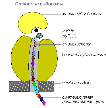
Ribosome structure
Ribosomes are synthesized in the nucleus, then leave it, moving into the cytoplasm, where they are attached to the outer surface of the membranes of the endoplasmic reticulum or are located freely. Depending on the type of protein being synthesized, ribosomes can function alone or be combined into complexes - polyribosomes.